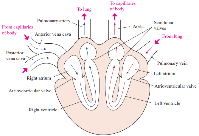
• Keep blood moving from ventricle and to arteries (aorta, pulmonary artery)
• Prevent backflow into ventricle
• Flap composed of connective tissue
Intercalated disks
• Contain gap junctions and mechanical junctions between cardiac muscle cells
• Allow electrical coupling and mechanical attachment between neighboring cardiac muscle cells
Atrium
• Chamber for receiving blood
Ventricle
• Chamber for pumping blood out
Conduction system
Sino-atrial node (SA node)
• Functions as pacemaker
• Located in the wall of the right atrium
• Initiates the heartbeat; conduction pathways coordinate atrial followed by ventricular contraction
Atrioventricular node (AV node)
• Located in the wall between right atrium and right ventricle
• Delays conduction to allow ventricular filling before ventricular contraction
c. Cardiac pressure
Systole – phase of ventricular contraction
Diastole – phase of ventricular relaxation
Cardiac output
– volume of blood pumped by a ventricle per minute; the left ventricle ejects into the aorta
– stroke volume × heart rate
Stroke volume
– volume of blood ejected by a ventricle per beat
3. Blood vessels
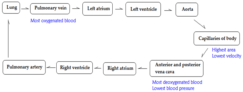
a. Blood circulation in the blood vessels
b. Functions of each part
Blood vessels
Features and functions
Artery
• Thick muscular wall and large vessel
• Transports blood from heart to arteriole
• Highest pressure
Arteriole
• Small vessel
• Transports blood from artery to capillary
• Branched from artery
• Determines the distribution of blood
Capillary
• Thin and smallest vessels with only endothelium tissues
• Highest area and lowest blood velocity
• Transports blood from arteriole to each body tissue
• Exchanges gases, nutrients, and wastes to body cells
Venule
• Thin and small vessel
• Collects blood from capillary
• The movements of skeletal muscles help return blood from capillary to heart in venule and vein
Vein
• Large vessel with low blood pressure
• Transports blood from venules to heart
• Has valves to prevent back flow
** Portal venous system
Transports blood from the capillaries to other capillaries via veins
Eg/ hepatic portal vein
c. Blood pressure
Main force for the circulatory blood movement
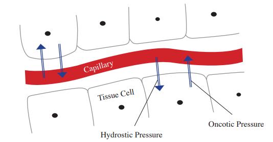
d. Starling forces
Driving forces for the fluid movement into and out of capillaries
Capillary hydrostatic pressure and capillary oncotic pressure
Starling forces
Features and functions
Capillary hydrostatic pressure
• Driving force of fluid from capillary to interstitial fluid.
• Net fluid movement depends on opposing hydrostatic and oncotic pressures and capillary permeability.
Capillary oncotic pressure
(Osmotic pressure)
• Driving force of fluid from interstitial fluid into capillary.
• Proteins of blood plasma in capillary pull water from interstitial fluid.
• The classical model predicts venous-end reabsorption; in many tissues at steady state, net filtration persists and lymphatics return the filtered fluid.
4. Blood
a. Cellular elements (45 % of blood)
Cell types
Features and functions
Erythrocytes
(Red blood cells)
• Have hemoglobin to transport oxygen and carbon dioxide
• Mature mammalian red blood cells lack nuclei and mitochondria
Leukocytes (White blood cells)
- cells of immune system
- have nuclei
- larger than red blood cells
- can move into the lymphatic system from the blood vessels
Basophils
• Secrete chemicals (histamine and anticoagulant heparin) to allergic reactions or damaged tissues
Eosinophils
• Minor phagocytic activity
• Release enzymes to kill parasites
• Sometimes discharges toxins from enzyme damage to tissues
Lymphocytes
• Helper T cells – secrete cytokines to activate B cells and cytotoxic T cells
• B cells – form plasma cells to secrete antibodies
• Cytotoxic T cells – secrete molecules (perforin) to kill infected or tumor cells
• Natural killer cells (Null cells)
- release factors to kill infected or tumor cells
Monocytes
• Become macrophages for phagocytic activity
Neutrophils
• Phagocytosis of microorganism or foreign particles
Platelets
• Hemostasis - process of blood clotting
• Lack nuclei
b. Plasma elements (55 % of blood)
Classification
Features and functions
Ion
• Regulation of osmotic balance, controlling of membrane permeability, pH buffering
– consists of a tetrameric protein in red blood cells.
– an allosteric protein.
– each subunit protein has a heme group, which contains iron.
– heme group binds O2 to transport.
– binding affinity of CO is greater than O2
** Myoglobin
– bind O2 to store in muscles.
– similar structure with hemoglobin but with monomer shape.
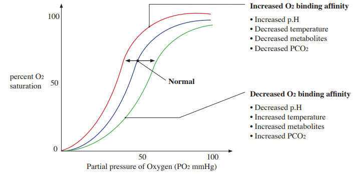
Oxygen – binding curve of hemoglobin
– increased O2 binding affinity means increased percent O2 saturation at the same partial pressure of O2
– cooperativity with oxygen binding to hemoglobin.
– Bohr effect means that O2 binding affinity of hemoglobin decreases with low pH (high CO2).
d. Transport carbon dioxide
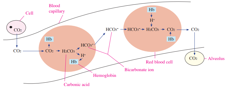
Most CO2 (70%) from respiring cells is transported to lungs as bicarbonate ion (HCO3–) mainly in plasma after bicarbonate is formed in red blood cells
Small amount of CO2 (23%) is bound to hemoglobin and transported
The rest of CO2 (7%) is dissolved in plasma
Blood pH is buffered in part by the carbonic acid–bicarbonate system (HCO3–)
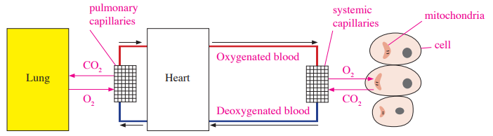
Exchanging O2 and CO2 by passive diffusion
e. States of red blood cells
f. Blood clotting
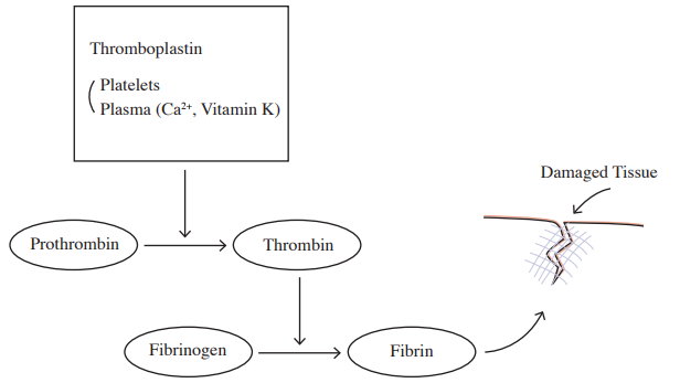
① The coagulation cascade, involving clotting factors, Ca2+, and platelet surfaces, converts prothrombin to thrombin. Vitamin K supports hepatic synthesis of several clotting factors.
② Thrombin enzyme converts an inactive fibrinogen to fibrin.
③ Fibrin forms framework of clot at damaged tissue.
Correction to the simplified sequence: red blood cells are produced in red bone marrow and circulate for about 120 days. Aged cells are removed by macrophages mainly in the spleen and liver; destruction is not confined to the liver.
C. Digestion
1. Mouth
Mechanical breakdown of food by teeth.
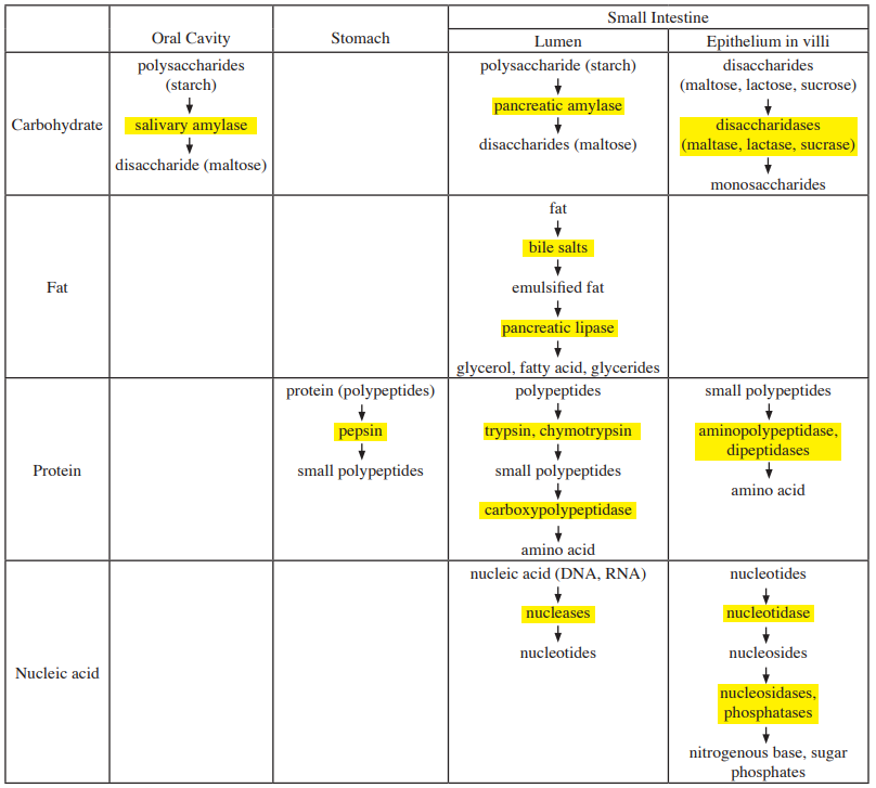
Chemical breakdown by salivary amylase, which breaks starch into maltose and shorter carbohydrate fragments.
Moistening of food to form a bolus.
2. Esophagus
Conducts food from mouth to stomach by waves of muscular contraction called peristalsis.
Controls the passage of food by muscular ring valves called sphincters.
3. Stomach
Functions for storage and digestion.
Mechanical breakdown by churning movements.
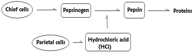
Gastric glands secrete mucus from mucous cells, pepsinogen from chief cells, and hydrochloric acid (HCl) from parietal cells.
Pepsin, which is converted from pepsinogen by HCl, digests proteins.
This is a simplified coagulation sequence. Vitamin K supports clotting-factor synthesis in the liver; it is not a component that directly converts prothrombin at the wound. The coagulation cascade generates thrombin, which converts fibrinogen to fibrin.
Food mixed with gastric juice becomes acid chyme.
Mucus lubricates and protects the surface of stomach walls.
Cardiac orifice (Gastroesophageal sphincter) regulates back flow of food from stomach to esophagus.
Pyloric sphincter controls the passage of acid chyme from stomach to small intestine.
4. Small intestine
: most chemical digestion for all nutrients occurs in small intestine, which is composed of duodenum, jejunum, and ileum.
a. Secretion to duodenum
Pancreas – secretes pancreatic juice, which contains the digestive enzymes and bicarbonate for neutralizing the acid chyme, to duodenum.
Liver – secretes bile, which contains bile salts that emulsify fats and aid their digestion, to duodenum.
Epithelium of small intestine – secretes digestive enzyme.
b. Absorption
Villi – highly folded mucosal projection to increase surface area for absorption.
Microvilli – microscopic projection on individual cells.
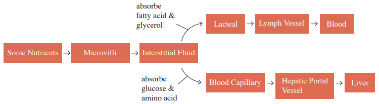
Long-chain fatty acids and monoglycerides enter intestinal epithelial cells, are reassembled into triglycerides, and leave in chylomicrons that enter lacteals. Short-chain fatty acids enter portal blood directly.
Glucose and amino acid are absorbed by blood capillary and carried to liver via hepatic portal vessel.
Correction to the lipid pathway: long-chain fatty acids and monoglycerides enter enterocytes, are reassembled into triglycerides, and are packaged in chylomicrons before entering lacteals. Short-chain fatty acids and glycerol can enter portal blood directly.
5. Liver
a. Secretion for digestion
Bile production and storage in gallbladder.
Bile contains bile salts and bicarbonate for helping lipid digestion as a function of the emulsifying agent but does not contain enzyme.
b. Multiple functions
Destruction of aged red blood cells.
Glycogen synthesis, breakdown, and storage.
Synthesis of plasma proteins such as albumin, clotting proteins, and angiotensinogen.
Conversion of ammonia to urea for excretion.
Elimination of waste or bacteria from blood.
6. Pancreas
a. Exocrine (secretion of pancreatic juice into the duodenum)
Secrete amylase to digest carbohydrate.
Secretes trypsinogen, which is activated to trypsin in the small intestine to digest proteins.
Secrete lipase to digest fat.
Secrete bicarbonate ions to neutralize the acid chyme.
b. Endocrine (secretion of hormones into the bloodstream)
α cells secrete glucagon to convert glycogen to glucose.
β cells secrete insulin to convert glucose to glycogen.
7. Large intestine (Colon)
Composed of four major parts, ascending colon, transverse colon, descending colon, and sigmoid.
Absorption of water, ions, and other minerals.
Feces transformation and storage of feces.
Absorption of vitamin K produced by resident gut bacteria.
8. Overview of digestion
Corrections to the digestion summary: pancreatic lipase mainly forms free fatty acids and monoglycerides from triglycerides. The protease labeled “carboxypolypeptidase” is carboxypeptidase. Bile salts emulsify fat rather than enzymatically hydrolyzing it.
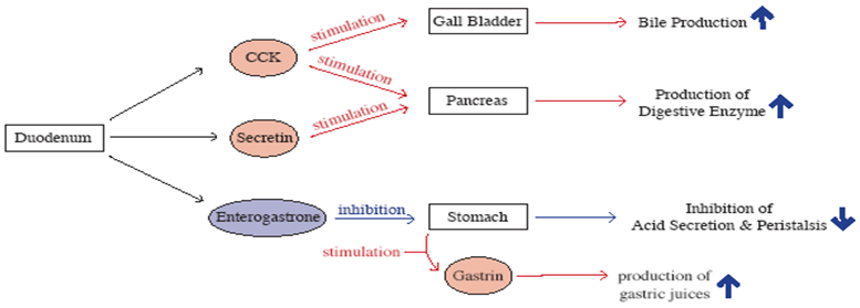
9. Hormonal control of digestion
Corrections to hormone targets: CCK promotes gallbladder contraction and pancreatic enzyme secretion; the liver produces bile. Secretin chiefly promotes bicarbonate-rich pancreatic and biliary secretion. Gastrin is secreted mainly by stomach G cells. “Enterogastrone” is a historical collective term for intestinal signals that inhibit gastric activity.
D. Osmoregulation and excretion
Osmoregulation – regulates the concentration of water and solute.
Excretion – eliminates the nitrogen containing waste product from metabolism.
1. Animal osmoregulation and excretion
Classification of animal
Osmoregulation and excretion
Flatworms
• Protonephridia with flame cells
Earthworm
• Metanephridia
Insect
• Malpighian tubules
Bony fish
Freshwater fish
• Excreting a large amount of water by kidneys for water balance in the hypotonic environment
Saltwater fish
• Marine bony fish drink seawater and excrete excess salts mainly through gill ionocytes; kidneys also eliminate some ions
Bird
• Marine birds use specialized salt glands to excrete excess salt; kidney function also contributes to water balance
2. Mammal osmoregulation and excretion
a. Basic renal processes of mammal kidney
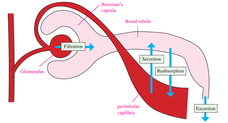
Filtration – hydrostatic pressure drives water and small solutes from glomerular blood into Bowman’s capsule; blood cells and most plasma proteins are retained.
Secretion – transport toxins and excess salts from body fluids (peritubular capillaries) to renal tubules by active transport.
Reabsorption – transport filtrated substances from renal tubules to body fluids (peritubular capillaries) by active or passive transports.
Excretion – elimination of urine from the body; excreted amount = filtered amount − reabsorbed amount + secreted amount.
• Glucose in protein-free plasma is filtered completely
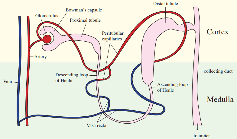
Bowman’s capsule
• Cup shaped renal tubes
• Surround glomerulus
• Filter water glucose and small solutes (sodium and potassium ions, amino acids, urea)
• Cannot filter large molecules such as blood cells or plasma proteins
Secretion (active transport)
and
Reabsorption ( active or passive
transport)
Proximal tubule
• Secretion and reabsorption of salt, nutrients (glucose and amino acid), and water
• Most of the reabsorption of water and salt occurs
• pH regulation
Distal tubule
• Secretion and reabsorption of salt and water
• Control K+ and NaCl
• pH regulation
• Aldosterone increases Na+ reabsorption and K+ secretion; water follows osmotically when the epithelium is water-permeable
Reabsorption ( active or passive
transport)
Descending loop of Henle
• Reabsorption of water
Ascending loop of Henle
• Reabsorption of NaCl
Excretion
Collecting duct
• Collects tubular fluid; ADH regulates water reabsorption and urine concentration
c. Passage of waste products
“Collection duct” should read collecting duct. After the collecting ducts, urine passes through the calyces and renal pelvis before entering the ureter; the diagram omits these intermediate structures.
E. Endocrinology
1. Hormone
: intercellular chemical signals produced by endocrine glands.
a. Hypothalamus hormones
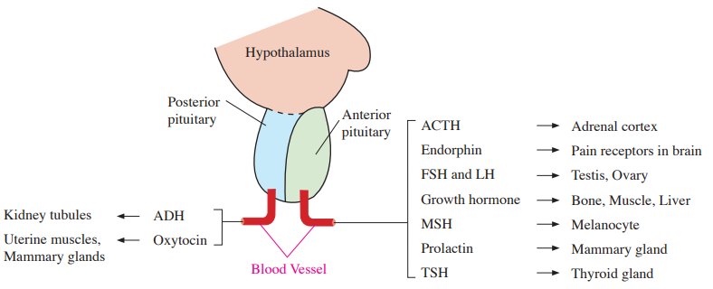
Hypothalamic releasing and inhibiting hormones regulate the anterior pituitary through portal blood. ADH and oxytocin are synthesized in the hypothalamus and released from the posterior pituitary.
b. Steroid hormones
Lipid-soluble and poorly soluble in water.
Cross plasma membranes and bind intracellular receptors in the cytoplasm or nucleus.
Cannot cross plasma membranes and bind to receptors on the surface of a target cell.
Eg/ Most hormones
d. Tropic hormones
Act on another endocrine gland to regulate its hormone secretion.
Eg/ FSH, LH, TSH, ACTH, hypothalamus hormones
e. Nontropic hormones
Eg/ Endorphin, MSH, prolactin
2. Hormones of endocrine glands
ADH and oxytocin are synthesized in hypothalamic neurons, transported down their axons, and released from the posterior pituitary. The posterior pituitary does not synthesize these hormones.
Hypothalamus Hormones
Pituitary
Hormone
Target
Functions of pituitary hormones
GHRH (stimulates GH)
GHIH
(inhibits GH)
Anterior Pituitary
GH
(growth hormone)
Liver and
bone
• Stimulates cell division and increasing cell size
TRH
(stimulates TSH)
TSH
(thyroid stimulating hormone)
Thyroid
• Controls homeostatic functions and cellular metabolism
• Primarily stimulates glucocorticoid secretion; aldosterone is mainly regulated by angiotensin II and plasma K+
GnRH
(stimulates LH and FSH)
LH (luteinizing hormone)
Testis and
ovary
• Causes the follicle to undergo ovulation
• Stimulates the Leydig cells for testosterone secretion in testes
FSH
(follicle stimulating hormone)
• Stimulates ovaries for primary follicle development
• Stimulates the Sertoli cells for spermatogenesis in testes
PRH
(stimulation) PIH
(inhibition)
Prolactin
Mammary
glands
• Stimulates mammary glands for milk production and secretion
MSH
Melanocytes
• Stimulates color changes of skin in reptiles and amphibians
Posterior
Pituitary
ADH
(antidiuretic hormone, vasopressin)
Kidney
tubules
• Stimulates the kidney for retention of water from renal tubules to body capillaries and increases blood pressure
• Causes concentrated urine
• Insufficient ADH causes Diabetes insipidus
Oxytocin
Mammary
glands,
uterine muscles
• Initiates milk release
• Induces labor
3. T3 and T4 from Thyroid Gland
: thyroid gland consists of two lobes sited on trachea and controls homeostatic functions and cellular metabolism such as digestion, heart rate, blood pressure, and reproductive functions.
Hypothalamic TRH stimulates pituitary TSH, which stimulates thyroid secretion of T3 and T4
Triiodothyronine (T3)
– has three iodine atoms.
– more powerful activation than T4.
Thyroxine (T4)
– has four iodine atoms.
– higher rate of production than T3.
– converted to T3 in kidney, liver, or target cells.
Metabolic disorders of the thyroid gland
Metabolic disorders
Causes and symptoms
Hyperthyroidism
• Excessive secretion of thyroid hormone
• High rate of metabolism with high temperature, poor emotional control, high blood pressure, irritability, profuse sweating, and weight loss
• Eg/ Graves’ disease (protruding eyes)
Hypothyroidism
• Insufficient secretion of thyroid hormone
• Low rate of metabolism with cold intolerance, lethargy, and weight gain
• Eg/ Myxedema, Cretinism in infants
4. Regulation of blood calcium levels
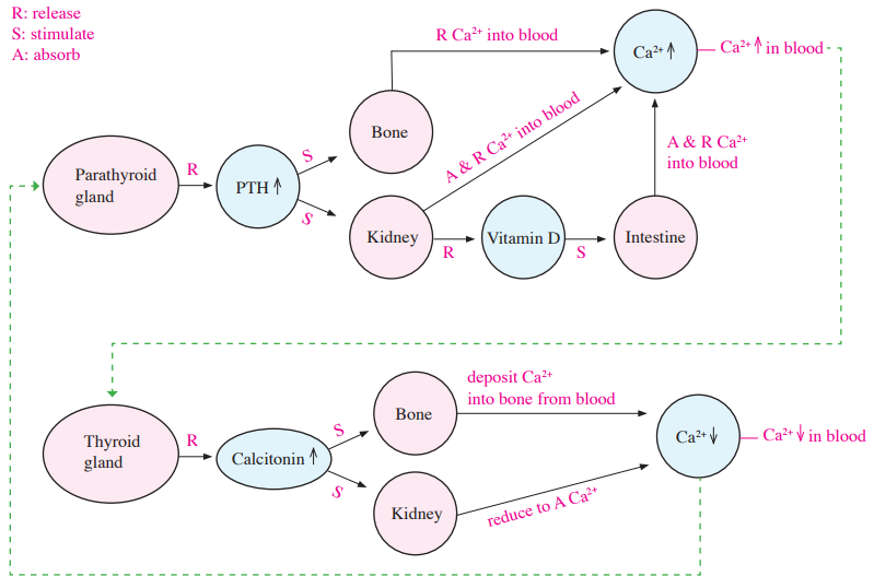
a. Parathyroid hormone (PTH)
Secreted by parathyroid gland when Ca2+ levels in blood are decreased.
Raises blood calcium by indirectly stimulating osteoclast-mediated bone resorption.
Increases renal calcium reabsorption and activates vitamin D, which increases intestinal calcium absorption.
b. Calcitonin
Secreted by thyroid gland when Ca2+ level in blood is increased.
Can lower blood calcium by inhibiting osteoclast-mediated bone resorption; it has a limited role in normal adult calcium homeostasis.
Can increase renal calcium excretion.
5. Regulation of blood glucose levels
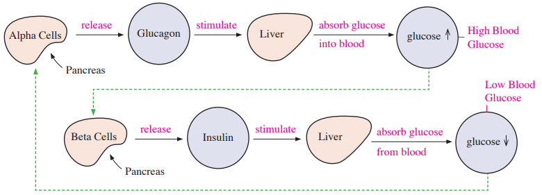
a. Glucagon
Secreted by alpha cells in pancreas when glucose levels in blood are decreased.
Conversion of glycogen to glucose.
Stimulates liver to release glucose into blood stream.
b. Insulin
Secreted by beta cells in pancreas when glucose levels in blood are increased.
Lowers blood glucose by promoting glucose uptake in skeletal muscle and adipose tissue, promoting glycogen synthesis, and suppressing hepatic glucose production.
Conversion of glucose to glycogen.
** Islet of Langerhans: composed of Alpha Cells, Beta cells, Delta cells, and F cells in pancreas
6. Stress response
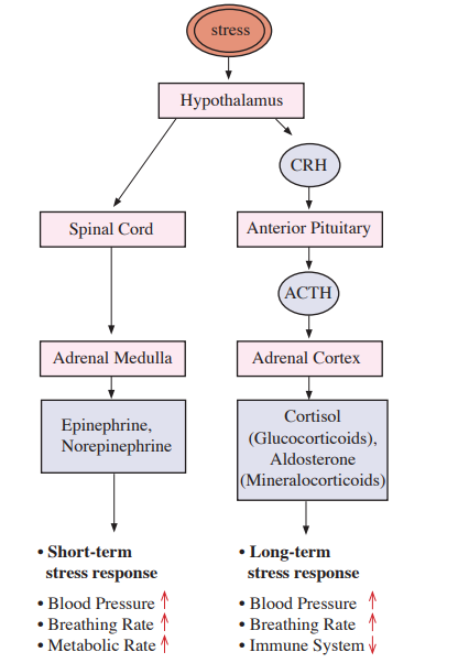
Regulated in part by adrenal glands located above the kidneys
Correction to the ACTH branch: ACTH mainly stimulates cortisol secretion. Aldosterone is controlled primarily by the renin–angiotensin system and plasma K⁺. Cortisol increases glucose availability and suppresses inflammation; increased breathing rate is not a defining direct cortisol effect.
a. Adrenal medulla
Stimulated from hypothalamus via spinal cord.
Secretes epinephrine and norepinephrine to response short term stress.
Raises blood glucose, heart rate, and blood pressure during the acute stress response.
b. Adrenal cortex
ACTH primarily stimulates glucocorticoid secretion; aldosterone is mainly regulated by angiotensin II and elevated plasma K+.