V. Genetics
Key focus of this chapter: DNA and Chromosome
This chapter reviews DNA, chromosomes, cell division, Mendelian genetics, protein synthesis, and mutation.
A. Cell Division
1. Cell cycle
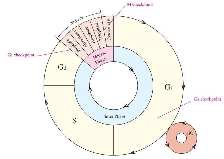
a. Controlling cell cycle
- G1 checkpoint
– determines whether cell cycle goes to S phase or the nondividing G0 state.
– cell growth checkpoint
- G2 checkpoint
– checkpoint before M phase.
– Checks completion of DNA replication and DNA damage.
– MPF is a cyclin–CDK complex that promotes entry into mitosis.
- M checkpoint
– Checks that all kinetochores are properly attached to spindle microtubules before anaphase.

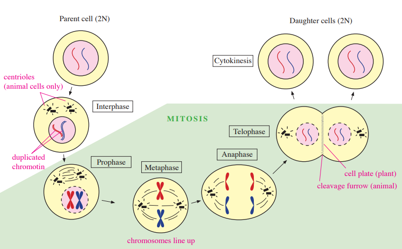
b. Mitosis
- Typically produces genetically identical daughter cells while preserving ploidy (e.g., 2n → 2n in diploid somatic cells).
- Occurs for repairing tissues and growing organisms.
Cell phases | Classification | Descriptions | |
Interphase | G1 phase | • Cell growth • First gap • Duration varies by cell type | |
S phase | • DNA synthesized • Duplicated chromatin | ||
G2 phase | • Cell growth and preparing for mitosis • Second gap | ||
M phase | Mitosis | Prophase | • Nuclear envelope disappears • DNA condenses into chromosomes • Spindle microtubule begins to form • Centrosomes move into opposite ends of the cell |
Metaphase | • Chromosomes align along the metaphase plate • Spindle fibers attach to the kinetochores | ||
Anaphase | • Separating and moving sister chromatids to opposite poles by spindle fibers | ||
Telophase | • Nuclear envelopes reappear • Chromosomes begin to be less condensed | ||
Cytokinesis | Cytokinesis | • Two daughter cells form • Plant cell: forms cell plate • Animal cell: forms cleavage furrow | |
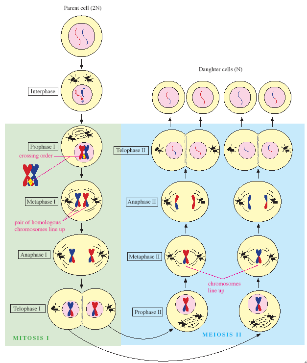
c. Meiosis
- Sexual reproduction resulting in spores or gametes.
- Reducing chromosome sets from diploid to haploid (2n → n) in germ line cells.
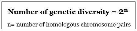
- Independent assortment and crossing over increase genetic diversity.
** DNA replication occurs during interphase before mitosis or meiosis I; it does not occur between meiosis I and II.
Meiosis I | Prophase I | • Nuclear envelope disappears • DNA condenses into chromosomes and then forms homologous chromosome pairs (tetrad) • Synapsis and crossing over of homologous chromosomes at chiasma • Spindle microtubule begins to form • Centrosomes move into opposite ends of the cell |
Metaphase I | • Pairs of homologous chromosomes line up along the metaphase plate • Spindle fibers attach to the kinetochores | |
Anaphase I | • Pairs of chromosomes separate and move to opposite poles by spindle fibers. | |
Telophase I & Cytokinesis | • Forming two haploid daughter cells • Plant cell: forming cell plate • Animal cell: forming cleavage furrow | |
Meiosis II | Prophase II | • Spindle microtubule begins to reform • Centrosomes move into opposite ends of the cell |
Metaphase II | • Chromosomes align along the metaphase plate • Spindle fibers attach to the kinetochores | |
Anaphase II | • Chromosome separate and sister chromatids move to opposite poles by spindle fibers | |
Telophase II & Cytokinesis | • Nuclear envelopes reappear • Chromosomes begin to be less condensed • Haploid daughter cells form • Cell plate of plant cell and cleavage furrow of animal cell |

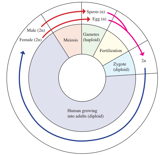
2. Cycle of sexual life
- Gametes
– reproductive germ-line cells
– haploid cells (n)
– eg/ Sperm and eggs
- Zygote
– fertilized egg
– diploid cell (2n)
- Somatic cells
– forming body of eukaryotic species except reproductive germ-line cell
B. Population genetics
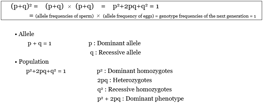
1. Hardy-Weinberg equilibrium
a. Equation
![]() |
b. Condition
- Large population
- Isolation (no gene flow)
- No mutation
- Random mating
- No natural selection
2. Genetic Drift
: random changes in allele frequencies, especially in small populations.
a. Bottleneck effect
- A sharp reduction in population size can change allele frequencies by chance.
b. Founder effect
- A small founding group may have allele frequencies unlike the source population and reduced diversity.
3. Genotype Frequency
a. Inbreeding
- Mating between relatives increases offspring homozygosity; it does not by itself change allele frequencies.
b. Hybridization
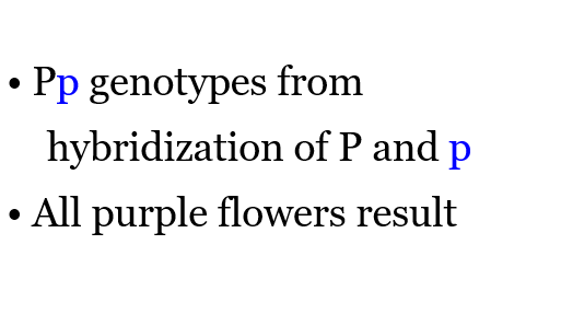
- Crossing genetically distinct individuals or varieties can combine parental alleles; outcomes vary.
C. Mendelian genetics
1. Terminology
- Allele
– alternative form of a gene.
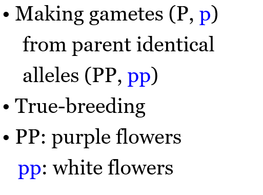
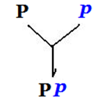
– eg/ P: dominant allele, p: recessive allele
- Phenotype: observable trait
- Genotype: genetic constitution
- Dominant allele
– expressed in a heterozygote under complete dominance.
– the dominant phenotype appears in dominant homozygotes and heterozygotes.
- Recessive allele
– masked by the dominant allele in a heterozygote under complete dominance.
-only appearance of the traits from recessive homozygotes.
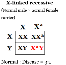
- Hemizygous – having one copy of a locus, such as most X-linked loci in an XY male.
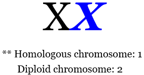
- Pair of homologous chromosomes.
– homologs carry the same gene loci, typically have similar size and centromere position, and may carry different alleles. One homologous pair contains two chromosomes; the image’s “1” means one pair, not one chromosome.
- Homozygous: A pair of identical alleles.
- Heterozygous: A pair of different alleles.
Pair of homologous chromosomes | Homozygous | Heterozygous |
![]() | AA, aa | Aa |
- Law of segregation
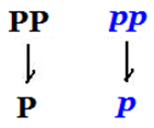
– the two alleles at a locus segregate so each gamete receives one.
- Law of independent assortment
– alleles at unlinked loci assort independently during gamete formation; closely linked loci may not.
- Testcross
– cross an individual with a dominant phenotype and unknown genotype with a homozygous recessive individual.
– all dominant offspring are expected if the unknown parent is homozygous dominant; a small sample cannot prove this genotype.
– a heterozygous parent gives an expected 1:1 dominant-to-recessive phenotype ratio.
2. Mendelian experiments
Classification | Figures | Features |
P generation | ![]() | ![]() |
F1 generation | ![]() | ![]() |
F2 generation | ![]() | ![]() |
- Monohybrids
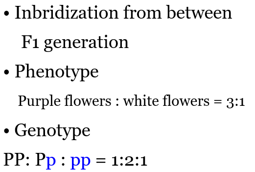
– crossing between two heterozygotes of F1 generation having a single characteristic.
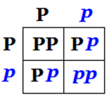
– eg/ Bb × Bb → Phenotypic ratio with 3:1
- Dihybrids
– crossing between two heterozygotes of F1 generation having two characteristics.
– eg/ BbDd × BbDd → An expected F2 phenotype ratio of 9:3:3:1 with independent assortment and complete dominance at both loci
3. Law of probability
Traits | Figures | Features and diseases |
Sex-linked traits | ![]() | • Passing the traits on X chromosome to offspring • For an X-linked recessive trait, sons are affected if they inherit the recessive allele from their mother • Daughters usually need a recessive allele from each parent; a carrier mother alone is insufficient. • Eg/ Hemophilia, color blindness |
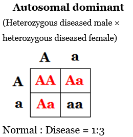
Autosomal traits | ![]() | • Dominant traits are expressed in heterozygotes and, when viable, dominant homozygotes • Eg/ Achondroplasia dwarfism, Huntington’s disease, progeria (premature aging) |
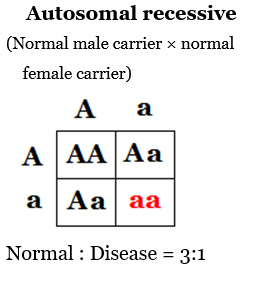
![]() | • Diseased offspring with homozygous recessive • Eg/ Sickle cell anemia, PKU disease, albinism, Galactosemia, Tay-Sachs disease |
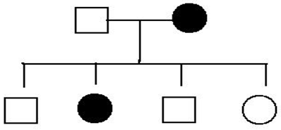
4. Pedigrees
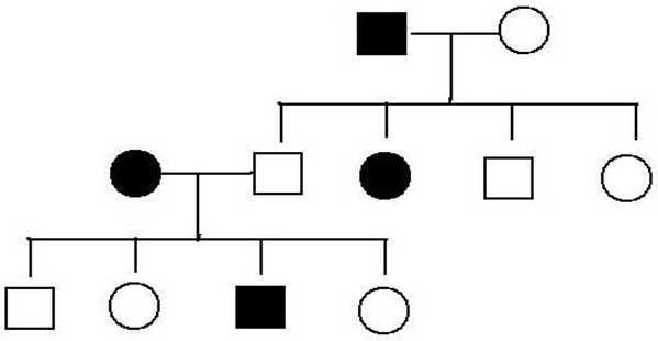
- Autosomal traits
– with full penetrance, an affected daughter and unaffected father exclude the usual X-linked recessive pattern; this observation alone does not prove autosomal inheritance.
- Dominant traits
– often appear in successive generations; reduced penetrance or a new mutation can alter this pattern.
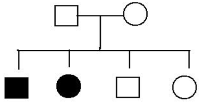
- Recessive traits
– unaffected carrier parents can have an affected offspring; recessive traits may skip generations.
5. Inheritance
a. Complete dominance
- The heterozygote has the same phenotype as the dominant homozygote.
- Purple flower × white flower → purple flower
b. Incomplete dominance
- The heterozygote has a phenotype intermediate between the two homozygotes.
- Eg/ White flower × red flower → pink flower
c. Codominance
- Both alleles are expressed in the heterozygote.
- Eg/ roan cattle: intermingled red and white hairs illustrate codominance
- Eg/ blood group (IA, IB, i), (multiple alleles or polymorphism)
Phenotype | Genotype | Antibodies |
A | ![]() | Anti-B |
B | ![]() | Anti-A |
AB | ![]() | None |
O | ![]() | Anti-A, Anti-B |
d. Pleiotropy
- One gene influences multiple phenotypic traits.
- Eg/ Sickle-cell disease
e. Epistasis
- One gene modifies or masks the phenotypic expression of another gene.
- Eg/ Color of mice
f. Polygenic inheritance
- Multiple genes contribute to a trait, often producing continuous variation.
- Eg/ Skin color
g. Penetrance
- The percentage of individuals with a given genotype who express its associated phenotype.
h. Multifactorial disorder
- A disorder influenced by both genetic and environmental factors.
i. Maternal effect
- The mother’s genotype determines offspring phenotype through products deposited in the egg. This differs from maternal mitochondrial inheritance.
D. Gene experiment
1. Griffith transforming experiment
a. Experiment
– 1) injection living S cells (encapsulated bacteria) into a mouse → mouse dies
– 2) injection living R cells (nonencapsulated bacteria) into a mouse → mouse lives
– 3) injection heat-killed S cells into a mouse → mouse lives
– 4) injection mixture of heat-killed S cells and living R cells into a mouse → mouse dies
b. Conclusion
- The living R bacteria were converted into the pathogenic S bacteria by an unidentified transforming factor.
2. Avery, MacLeod, and McCarty experiment
a. Experiment
– 1) mixture heat-killed S cells, living R cells, and protease → living S cells
– 2) mixture heat-killed S cells, living R cells, and ribonuclease → living S cells
– 3) mixture heat-killed S cells, living R cells, and deoxyribonuclease → no living S cells
b. Conclusion
- DNA is the transforming factor.
E. Chromosome basics
1. Chromosomes in humans
- 46 chromosomes in diploid cells (2n).
- 23 chromosomes in haploid gametes (n).
- Typically 44 autosomes and two sex chromosomes (usually XX or XY).
2. Chromosome appearances
Classification | Features |
Metacentric | Location of centromere at middle of chromosome |
Acrocentric | Location of centromere at close to end of chromosome |
Telocentric | Location of centromere at end of chromosome |
3. Level of chromosomal organization

- Histone: DNA packing molecules composed of protein.
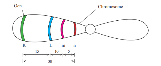
4. Linked genes
: genes on the same chromosome tend to be inherited together; crossing over can separate their alleles.
- Recombination frequency estimates genetic distance: 1% recombination ≈ 1 centimorgan for short intervals.
- Increased probability of crossing over with greater the gap between two linked genes.
- Linkage map
5. Sex-linked genes
: genes on X or Y chromosomes.
- Sex-linked recessive traits
– hemophilia
– color blindness
- Barr body: an inactivated x-chromosome
6. Chromosomal alteration
a. Changing chromosome number
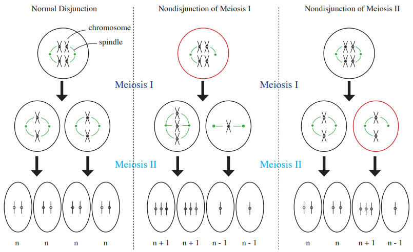
- Nondisjunction
– separation failure of pairs of homologous chromosomes in meiosis I or sister chromatids in meiosis II.
- Aneuploidy
: abnormal number of chromosomes from the nondisjunction of meiosis I or II.
– monosomy (2n − 1): one chromosome of a homologous pair is missing
– trisomy (2n + 1): three copies of one chromosome
- Polyploid
: more than two sets of chromosomes.
– triploidy (3n)
– tetraploid (4n)
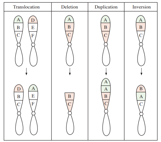
b. Changing chromosomal structures
- Translocation
– transfer of a chromosome segment to a nonhomologous chromosome.
- Deletion
– lost chromosomal segment.
- Duplication
– repeating chromosomal segment.
- Inversion
– reverse attachment of chromosomal segment.
7. Human disorders
a. Down syndrome
- Usually trisomy 21 from nondisjunction; translocation and mosaic forms also occur
- 47 chromosomes
b. Klinefelter syndrome
- Nondisjunction of X chromosome
- Usually an XXY male with impaired fertility
- May include gynecomastia and reduced testicular function
- 47 chromosomes
c. Turner syndrome
- Nondisjunction of X chromosome
- Usually a female with a 45,X karyotype and ovarian insufficiency; mosaic forms occur
- Monosomy of sex chromosome
- 45 chromosomes
F. DNA replication
1. DNA structure

- Rosalind Franklin made X-ray diffraction photograph.
- James Watson and Francis Crick proposed the DNA model of double helix structure.
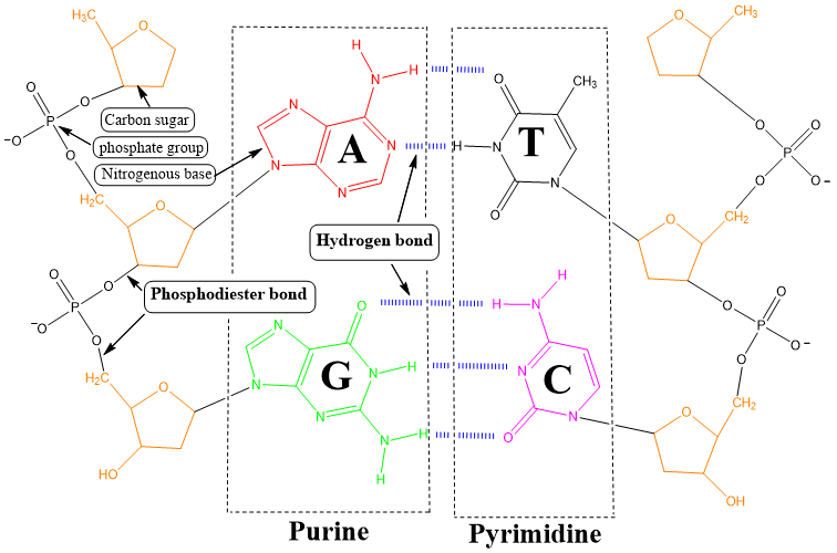
- DNA backbone: deoxyribose sugars bound to phosphate groups.
- Purine: A (adenine), G (guanine).
- Pyrimidine: C (cytosine), T (thymine).
- Two hydrogen bonds between A and T, three hydrogen bonds between G and C.
- With comparable length and conditions, higher GC content generally increases DNA melting temperature.
2. Chargaff’s rules
- A+G = C +T
- Eg/ A = 30%, G = 20%, C = 20%, T = 30%
3. Semiconservative model
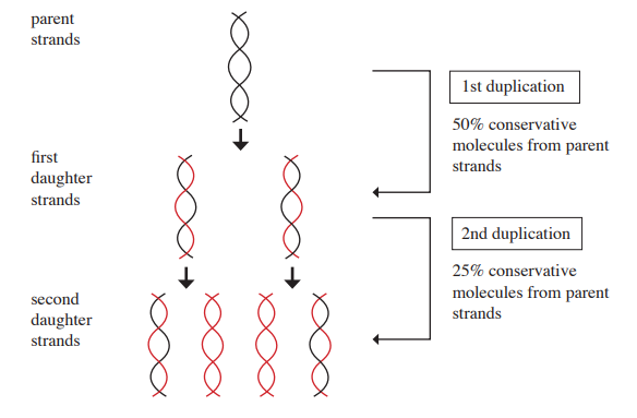
- The parental strands separate and each serves as a template for a new complementary strand.
- Each daughter DNA molecule contains one parental strand and one newly synthesized strand.
- After one replication in unlabeled medium, parental label constitutes half the total DNA if only parental DNA was labeled.

4. DNA replication
DNA replication | RNA base pairing |
A ⇔ T G ⇔ C | A ⇔U G ⇔ C |
a. Origins of replication
- Replication fork
– Y shaped site by helicases.
– places of elongation of new strand DNA.
b. Elongation
- Leading strand
– DNA Pol III elongates continuously new strand of DNA from 5´ to 3´.
– same direction with movement of replication fork.
- Lagging strand (Okazaki fragments)
– DNA Pol III creates short segments of new DNA from 5´ to 3´.
– opposite direction against movement of replication fork.
– each Okazaki fragment is synthesized 5′ → 3′; fragment formation proceeds away from the moving fork.
c. Enzymes for DNA synthesis
- Topoisomerase
– relieving the strain of tighter twisting from untwisting of parental DNA.
- Helicase: Separating the parental double strands of DNA
- Single-strand binding protein
– stabilizing single-stranded DNA and preventing reannealing.
- Primase
– synthesizing short segments of RNA primer which is nucleotide chain for starting point of DNA synthesis and putting down the RNA primer on new strands of DNA.
- DNA polymerase
– In bacteria, DNA Pol III adds nucleotides to extend DNA; eukaryotes use different replicative polymerases.
– In bacteria, DNA Pol I removes RNA primers and replaces them with DNA.
– proof reading (DNA error correction).
- DNA ligase
– joining the ends of new strand of DNA.
- Telomerase
– uses an RNA template to extend telomeres (TTAGGG repeats in humans), helping offset replication-associated shortening.
d. Repairing DNA
G. Making protein
1. Central dogma
2. Transcription
a. Initiation
① In eukaryotic RNA polymerase II transcription, general transcription factors assemble at the promoter; some promoters contain a TATA box.
② RNA polymerase binds to promoter along with the transcription factors.
③ RNA polymerase binding on DNA begins to synthesize pre-mRNA.
b. Elongation
- RNA Polymerase elongates pre-mRNA from 5´ to 3´
c. Termination
- Finishing pre-mRNA transcript
3. RNA processing
a. Modifying of pre-mRNA ends
- Adding 5´cap and poly-A tail to pre-mRNA ends
b. RNA splicing
: spliceosome removes introns from pre-mRNA and splices exons together to make mRNA.
- Introns: noncoding segments of nucleic acid
- Exons: sequences retained in mature RNA; they can include coding regions and untranslated regions
- Spliceosome: snRNPs + other proteins
4. Translation
a. Codons and anticodons
- Codons: triplet nucleotides at mRNA and translated into amino acids
– start codon: AUG (met)
– stop codon: UAA, UAG, UGA
- Anticodons: triplet nucleotides at the end of tRNA and complementary on codon
- Wobble: flexible pairing at the third base of a codon lets some tRNAs recognize multiple codons; this contributes to genetic-code degeneracy
b. tRNA
- Features
– hydrogen bonds with short double stranded RNA.
– presence of unusual bases: methylated bases, pseudouridine, 4-thiouridine.
- Aminoacyl-tRNA synthetase binds an amino acid to 3´ end of tRNA
c. Ribosomes
: synthesizes a polypeptide while reading mRNA 5′ → 3′; initiation, elongation, and termination use GTP.
- A site
– aminoacyl-tRNA binding site.
- P site
– holds the tRNA carrying the growing peptide. Peptidyl transferase transfers the peptide to the amino acid on the A-site tRNA; translocation then moves tRNAs through the sites.
- E site
– exit of tRNA site.
H. Mutation
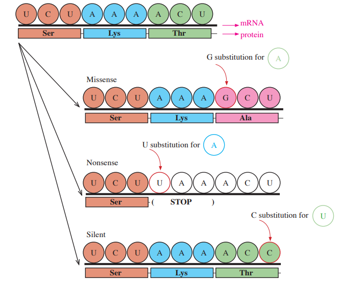
1. Point mutation
: substitution of one nucleotide molecule.
- Missense: result of a different amino acid by altering codon
- Nonsense: result of stop codon
- Silent: result of same amino acid although altering codon
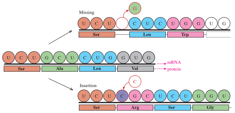
2. Frame shift
: insertion or deletion of a number of nucleotides not divisible by three within a coding sequence.
- Can disrupt downstream codons and protein function
- Missing or insertion
3. Cancer
- Oncogenes: genes leading to cancer.
- Cancer usually develops through accumulated changes in growth-control genes, including oncogenes and tumor-suppressor genes.
I. Gene technology
1. Polymerase chain reaction (PCR)
- Quickly copy and amplify an original double strand of DNA.
- Useful for critical forensic analysis.
2. X-ray diffraction
- Method to know the three-dimensional molecular structure.
- X-ray crystallography.
3. DNA fingerprint
- Identification of an individual’s DNA by their DNA profiles.
4. Transgenic organism
- An organism containing introduced genetic material from another source.