VII. Nervous system
Key focus of this chapter: neuron and nervous system
This chapter reviews neurons, neural signaling, and the central and peripheral nervous systems.
A. Organization of the nervous system
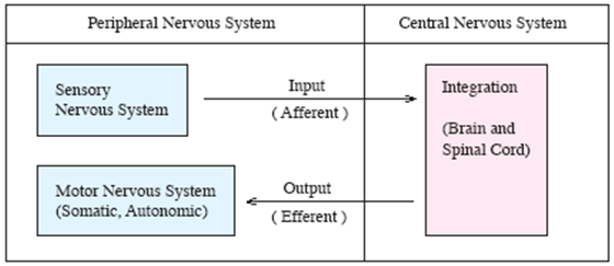
- Afferent
– carry sensory information toward the central nervous system.
- Interneurons
– process and integrate information within the central nervous system.
- Efferent
– carry motor commands away from the central nervous system to effector organs.
B. Structure of neuron
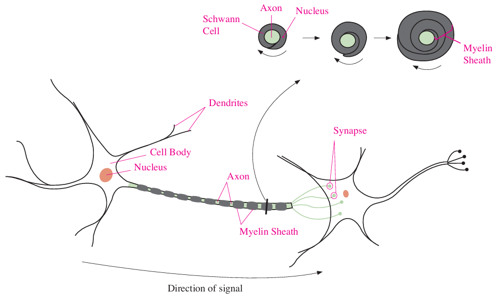
- Dendrites
– receive incoming signals.
– usually highly branched.
- Cell body
– contains the nucleus and supports cellular metabolism and protein synthesis.
- Axon
– conducts action potentials, normally from the initial segment toward the axon terminals.
- Myelin sheath
– Schwann cells form myelin around peripheral axons; oligodendrocytes form myelin in the central nervous system.
– myelin insulates axons and increases action-potential conduction speed.
** Synapse
: chemical synapses normally transmit from the presynaptic terminal to the postsynaptic cell; electrical synapses may transmit in both directions.
C. Transmission
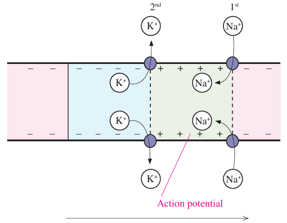
1. Transmission of action potential
- ① Voltage-gated Na+ channels open, allowing Na+ influx and depolarization.
- ② Na+ channels inactivate and voltage-gated K+ channels open; K+ efflux repolarizes the membrane.
- ③ Propagation of action potential.
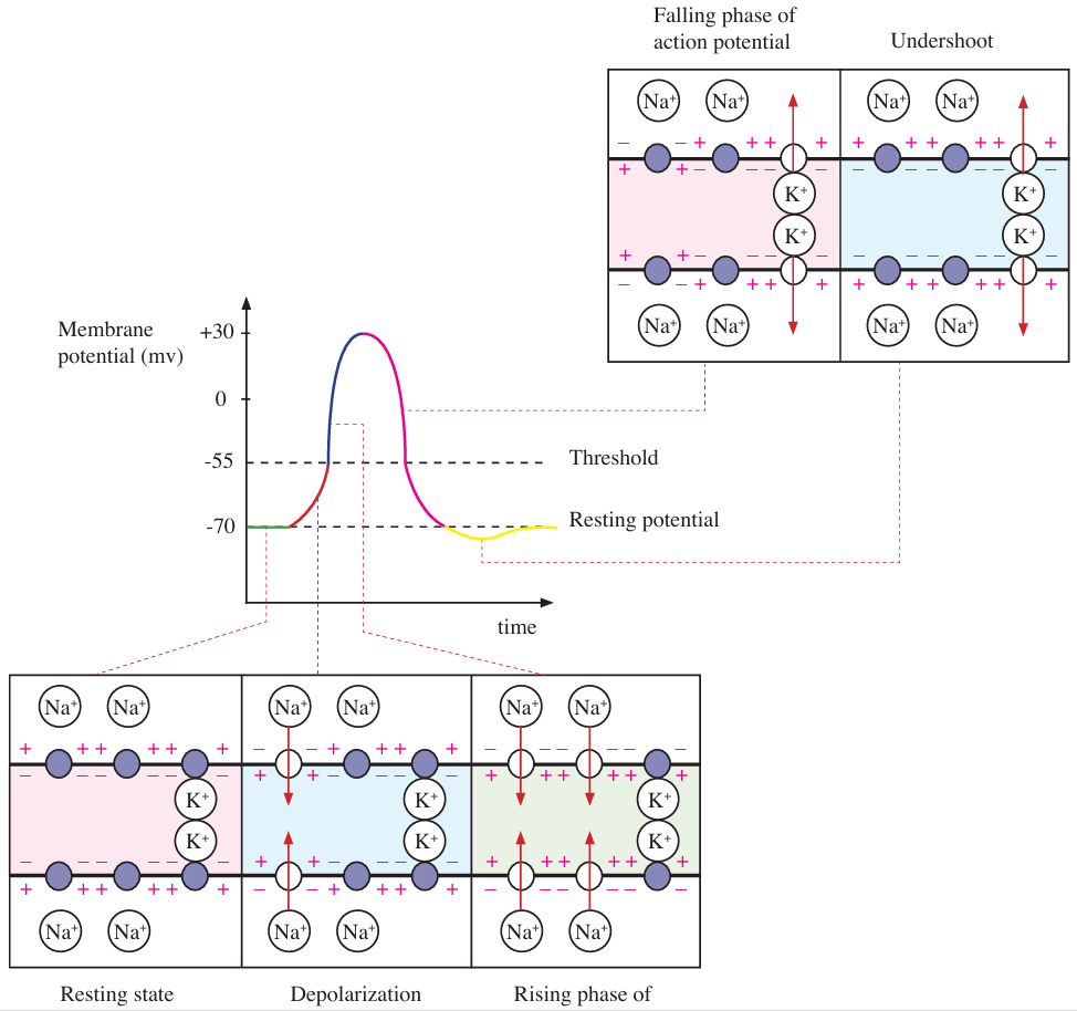
2. Action potential with voltage-gated ion channels

a. Resting state
- At rest, most voltage-gated Na+ and K+ channels are closed; leak channels remain open and the Na+/K+ pump maintains ion gradients.
- The cell interior is negative relative to the exterior; Na+ concentration is higher outside and K+ concentration is higher inside.
b. Depolarization
- Beginning depolarization and opening Na+ channels.
- Membrane depolarization reaches threshold.
c. Rising phase of action potential
- Increasing Na+ influx into inside of neuron with more opening of Na+ channels.
- Membrane potential rises to the action-potential peak.
d. Falling phase of action potential
- Voltage-gated Na+ channels inactivate while voltage-gated K+ channels open.
- K+ efflux makes the membrane potential more negative (repolarization).
e. Undershoot
- The membrane briefly becomes more negative than its resting potential because voltage-gated K+ channels close slowly; Na+ channels recover from inactivation.
- During the absolute refractory period, inactivated Na+ channels prevent another action potential; during the relative refractory period, a stronger stimulus is needed.
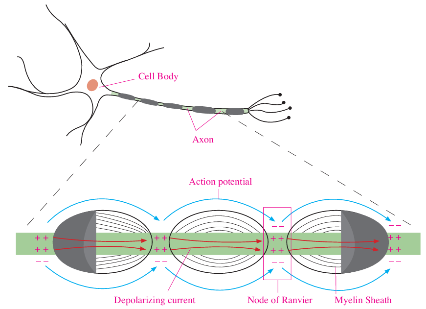
3. Saltatory conduction
: fast propagation of action potential along with a myelinated axon.
- Action potential jumps from node of Ranvier to next node of Ranvier along insulated myelin sheath.
- Myelination and larger axon diameter generally increase conduction speed; the number of myelinated axons does not determine speed in an individual axon.
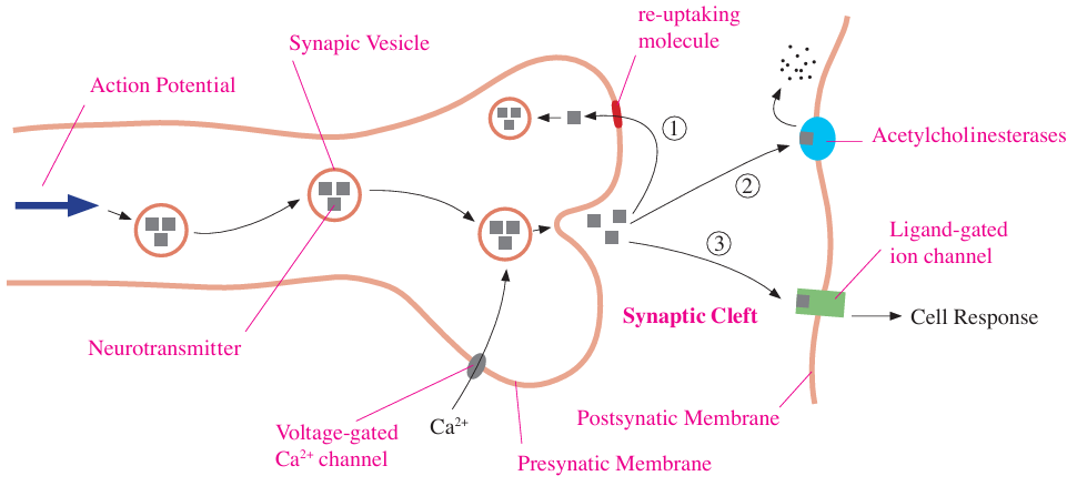
4. Chemical synapse
: place for transmission of messages between two neurons or between a neuron and a target cell.
a. Processing
- ① An action potential reaches the presynaptic terminal.
- ② Neurotransmitters in synaptic vesicles.
- ③ Voltage-gated Ca2+ channels open and Ca2+ enters the presynaptic terminal.
- ④ The rise in presynaptic Ca2+ triggers vesicle fusion and neurotransmitter release into the synaptic cleft.
- ⑤ Neurotransmitter binds postsynaptic receptors and produces an excitatory or inhibitory postsynaptic response. An action potential occurs only if excitation brings the axon initial segment to threshold.
- ⑥ Neurotransmitter is removed by reuptake, diffusion, or enzymatic breakdown; acetylcholinesterase specifically breaks down acetylcholine.
b. Function of each part
- Neurotransmitter
– intercellular chemical messengers.
- Synaptic vesicles
– packaging of neurotransmitter in presynaptic cell.
- Voltage-gated Ca2+ channel
– opening of Ca2+ channel when action potential reaches a synapse.
- Synaptic cleft
– narrow gap between presynaptic membrane and postsynaptic membrane.
– neurotransmitters diffuse across the cleft to postsynaptic receptors.
- Ligand-gated ion channels
– receptor of neurotransmitter at postsynaptic membrane.

D. Central nervous system
1. Brain
Regions | Classification | Functions and features | |
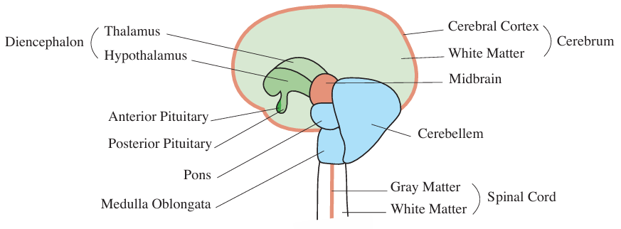
Forebrain | Cerebrum | • Integrating and analyzing sensory information, communication, language, learning, remembering, creative thought, motor response. • Consists of cerebral cortex (gray matter), white matter, and basal nuclei • Gray matter on the outside– dendrite, cell body • White matter on the inside-myelinated axon • Corpus callosum – communication between left brain and right brain | |
Diencephalon | Thalamus | • Sensory input and output for cerebrum • Control of sleeping and wakefulness | |
Hypothalamus | • Regulation of homeostasis such as blood pressure, temperature, or water balance • Sexual drive, thirst, hunger | ||
Midbrain | Midbrain | • Part of brainstem | |
Hindbrain | Pons | • Regulation of breathing rates | |
Medulla oblongata | • Regulation of cardiovascular and respiratory functions, swallowing, and digestive reflexes | ||
Cerebellum | • Coordination, equilibrium (motor balance) | ||
- Brainstem – midbrain, pons, medulla oblongata
- Meninges – membranes covering the central nervous system
2. Spinal cord
: communication of neural information between brain and body parts and regulation of reflexes.
- White matter on outside – transmission of neural information
- Gray matter on inside – processing of information
E. Peripheral nervous system
Peripheral nervous system | ||
Autonomic nervous system | Somatic nervous system | |
• Involuntary control • Autonomic efferent (motor) pathways respond to visceral sensory input response from internal stimuli such as smooth muscle, cardiac muscles, cardiovascular, digestive organs, excretory, and endocrine. | • Motor control of skeletal muscles, including voluntary movement and reflexes • Efferent (motor) nerves - response from external stimuli - reflex activity by spinal cord | |
Parasympathetic division | Sympathetic division | |
• Conserving energy and supporting rest-and-digest functions • Slows heart rate and promotes digestive activity; effects vary by target organ • Increasing digestion • Vagus nerve (cranial nerve X) supplies parasympathetic fibers to thoracic and many abdominal organs; pelvic splanchnic nerves supply the distal colon and pelvic organs | • Increasing energy generation from stress • Respond with increasing heart rate, respiration, rate of converting glycogen to glucose, and secretion of epinephrine • Decreasing digestion | |
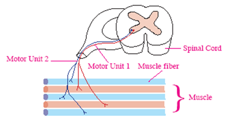
1. Motor unit
- Consists of one motor neuron and all skeletal muscle fibers it innervates.
- One neuron controls many muscle fibers.
2. Autonomic neurotransmitters and receptors
Exceptions: sympathetic postganglionic fibers to most sweat glands release acetylcholine onto muscarinic receptors. Preganglionic sympathetic fibers stimulate adrenal-medulla chromaffin cells through nicotinic receptors, releasing epinephrine and norepinephrine into the bloodstream.
Preganglionic neuron | Postganglionic neuron | |||
Neurotransmitter | Receptor | Neurotransmitter | Receptor | |
Parasympathetic system | ACh (Acetylcholine) | Nicotinic cholinergic | ACh | Muscarinic cholinergic |
Sympathetic system | ACh | Nicotinic cholinergic | NE (Norepinephrine) | Adrenergic |