VI. Embryology

Key focus of this chapter: male and female reproductive system

This chapter reviews reproductive systems, fertilization, embryonic development, extraembryonic membranes, and the mammalian placenta.

A. Male reproductive system

1. Anatomy

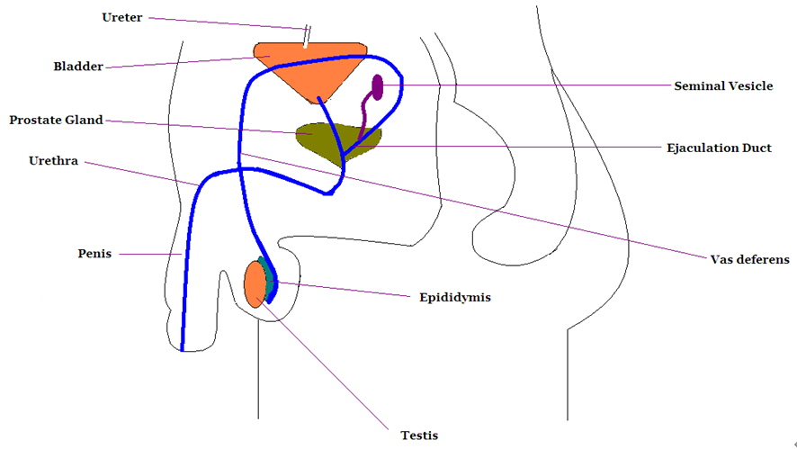
Male reproductive system

Functions

Testis

Seminiferous

Tubules

• Production of sperm

• Spermatogonia

Leydig cells (Interstitial cells)

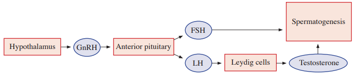
• Stimulated by luteinizing hormone (LH) to synthesize androgens, chiefly testosterone

Sertoli cell

• Nurturing and controlling sperm development

Epididymis

• Place for maturation and storage of sperm

Prostate gland

• Neutralizing the acidity of the vaginal tract and seminal fluid by secreting slightly alkaline milky fluid

• Enhancing motility and life of sperm

Seminal vesicle

• Secretes fructose-rich fluid that nourishes sperm and contributes to semen

Vas deferens

• Duct for expulsion of sperm

Ejaculatory duct

• Joining ducts of seminal vesicle and vas deferens

Urethra

• Duct of semen and urine

Scientific schematic
Leader-line correction: the urethra runs from the bladder through the prostate and penis. The vas deferens ascends from the epididymis and joins the seminal-vesicle duct to form the ejaculatory duct; the crossing leader lines in this schematic should not interchange these ducts.

2. The passage of sperm

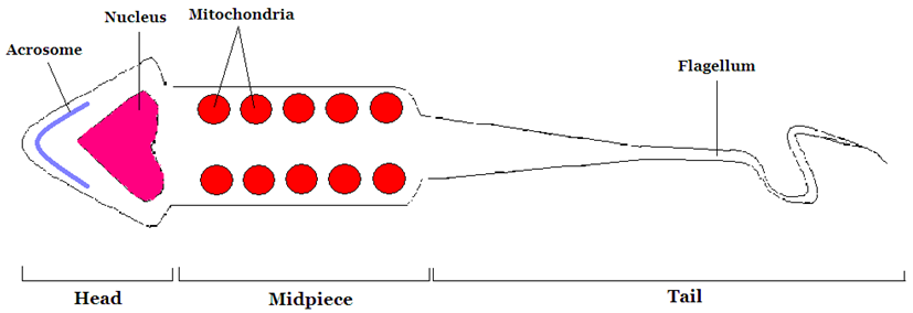
3. Sperm

  • Acrosome

– contains enzymes that help sperm penetrate the egg’s outer coverings during fertilization

  • Nucleus
  • Mitochondrion
  • Flagellum

– movement of sperm

4. Hormone regulation

B. Female reproductive system

1. Anatomy

Scientific schematic
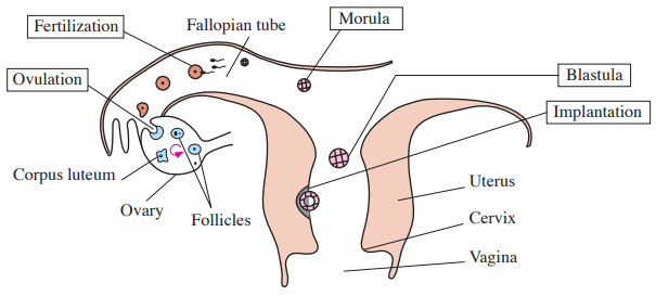
In mammals, the implantation-stage structure labeled “blastula” is a blastocyst. A secondary oocyte is ovulated; fertilization usually occurs in the ampulla of the uterine tube before cleavage and implantation.

Female reproductive system

Functions

Follicle

• An ovarian oocyte surrounded by supporting follicular cells

Ovary

• Place of oogenesis, which is the process of developing of mature ova

Ovulation

• A mature follicle releases a secondary oocyte, arrested in metaphase II

• Ruptured follicle becomes corpus luteum

Corpus luteum

• Temporary endocrine structure from the ruptured follicle; secretes progesterone and estrogens

Oviduct (Fallopian tube)

• Place of fertilization or passage of egg

Uterus

• Implantation of the blastocyst in the endometrium 

2. Passage of an ovum

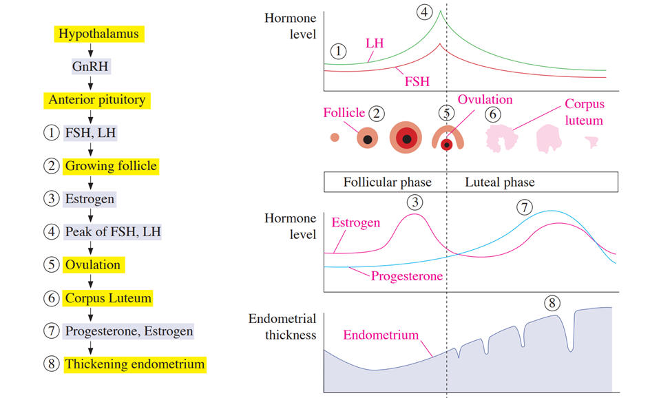
3. Hormone regulation

  • FSH

– supports spermatogenesis through Sertoli cells in males and follicular development in females.

– promotes growth of ovarian follicles.

  • LH

– stimulates testes and ovaries.

– LH surge causes follicle to undergo ovulation and convert into corpus luteum.

  • Estrogen

– prepares uterine lining growth for the implantation of the blastocyst.

– development of female secondary sex characteristics.

  • Progesterone

– maintains the secretory endometrium for the implantation of the blastocyst.

** Estrogen and progesterone usually provide negative feedback. Sustained high estrogen late in the follicular phase switches to positive feedback, producing the LH surge.

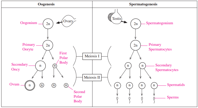
4. Comparison of oogenesis with spermatogenesis

Scientific schematic
Human timing: primary oocytes arrest in prophase I; the ovulated secondary oocyte arrests in metaphase II and completes meiosis II after fertilization. Four spermatids mature into sperm through spermiogenesis. The first polar body may divide, so polar-body number varies.

a. Reproduction of oogenesis

  • Produces one functional ovum and polar bodies
  • Unequal distribution of the cytoplasm

b. Reproduction of spermatogenesis

  • Produces four functional spermatozoa
  • Cytoplasm equally distributed

C. Fertilization

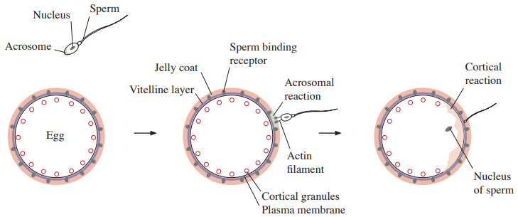
1. External fertilization of sea urchin

a. Process of fertilization

  • ① Contact – Sperm contacts the egg’s jelly coat.
  • ② Acrosomal reaction – Acrosomal enzymes digest the jelly coat; the acrosomal process binds receptors in the vitelline layer.
  • ③ Depolarization – Sperm–egg membrane fusion rapidly depolarizes the egg membrane, producing the fast block to polyspermy.
  • ④ Insertion of sperm nucleus into egg.
  • ⑤ Cortical reaction – Release of cortical-granule contents transforms the vitelline layer into fertilization envelope and causes a slow block to polyspermy.

b. Function of each part

Parts

Functions

Acrosome

• Contains hydrolytic enzymes that digest the sea-urchin egg’s jelly coat

Jelly coat

• Envelope of eggs

• Providing material for the sperm to contact on surface of eggs

Vitelline layer

• Extracellular matrix outside of plasma membrane and inside of jelly coat

• Place for sperm binding receptors

Sperm binding receptor

• Binding sperm head to contact at vitelline layer

Cortical granules

• Causing cortical reaction to block polyspermy

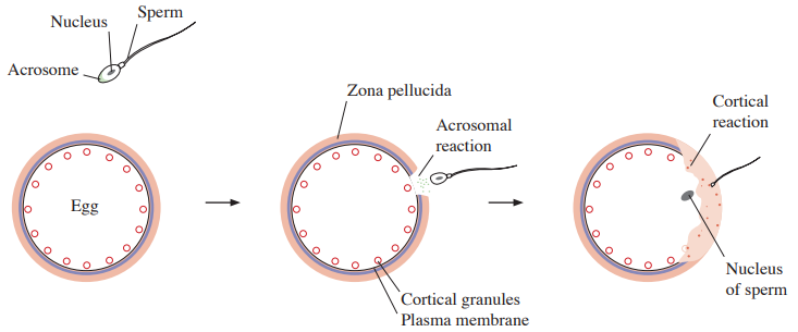
2. Internal fertilization of mammals

a. Process of fertilization

  • ① Approaching – sperm approach the zona pellucida of egg.
  • ② Acrosomal reaction – acrosomal enzymes and sperm motility help penetrate the zona pellucida, allowing sperm to reach the egg membrane.
  • ③ Sperm and egg plasma membranes fuse.
  • ④ Insertion of sperm nucleus into egg.
  • ⑤ Cortical reaction – release of cortical-granule contents modifies the zona pellucida and causes a slow block to polyspermy.

b. Function of each part

Parts

Functions

Acrosome

• Contains enzymes that help sperm penetrate the zona pellucida

Zona pellucida

• Extracellular matrix of egg

• Sperm receptor

Cortical granules

• Causing cortical reaction of slow block to polyspermy

c. Twins

  • Identical twins – one zygote splits into two embryos with essentially the same initial genotype
  • Fraternal twins – two eggs fertilized by separate sperm develop into two embryos

d. Cell differentiation

  • Becoming more specialized cell types in function and structure
  • Single zygote becomes specialized system of tissues in multicellular organism

D. Embryonic development

1. Description of each stage

Stages

Description

Cleavage
(Zygote cell division)

Morula

• Forming a solid ball of cells

Blastula

• Forming a hollow ball of cells
• Consists of blastocoel (fluid-filled cavity)

Gastrulation

• Cell movements establish germ layers; a dorsal blastopore lip forms in amphibian gastrulation

• Forming three germ layers (Ectoderm, mesoderm, endoderm) and primitive gut

• Shape and cell movements vary by animal group

• Archenteron – becomes primitive gut

• Blastopore
- protostome becomes mouth

- deuterostome becomes anus

• Involution – surface cells of embryo roll over into interior of embryo
Organogenesis

• Becoming different rudiments of organs from three germ layers

• Neurulation - developmental stages of notochord and hollow neural tube

• Induction – signaling from one tissue influences the developmental fate of another
• Forming neural crest, somites, notochord, neural tube

2. Three germ layers

Ectoderm

Mesoderm

Endoderm

• Epidermis of skin

• Sensory receptor of epidermis

• Nervous system

• Lens of eyes

• Tooth enamel

• Adrenal medulla

• Circulatory system

• Excretory system

• Reproductive system (except germ cells)

• Lymphatic system

• Connective tissue

 (Dermis of skin)

• Skeletal system

• Muscular system

• Adrenal cortex

• Epithelial lining of the respiratory tract

• Most digestive-tract epithelium; liver and pancreatic epithelium

• Most urinary-bladder epithelium

** Totipotent: a cell can generate all embryonic and extraembryonic cell types, potentially producing an entire organism

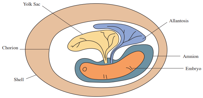
E. Functions of extraembryonic membranes in the shelled eggs

Classification

Functions and features

Amnion

• Membranous sac for protecting an embryo from mechanical shock and dehydration

• Not present in amphibians

Chorion

• Membrane for exchanging oxygen and carbon dioxide between embryo and outside membrane

Allantois

• Stores nitrogenous wastes and contributes to gas exchange in shelled eggs

• In mammals, associated tissues contribute to umbilical vessels; the chorion forms the fetal placental component

Yolk sac

• Provides nutrients to embryo

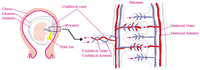
F. Placenta in mammal

: an exchange organ with fetal and maternal components; nutrients, gases, and wastes cross a placental barrier while the two blood supplies normally remain separate.

1. Placental circulation

: maternal placenta provides nutrients and oxygen to fetus and accepts waste product and carbon dioxide from fetus.

Scientific schematic
There is normally one umbilical vein and two umbilical arteries. The vein carries fetal blood from placenta to fetus; the arteries return fetal blood to placenta. Maternal blood bathes chorionic villi while fetal blood remains in villous vessels, so exchange normally occurs without direct mixing.

2. Function of fetal placenta

  • Umbilical vein – carries oxygenated, nutrient-rich blood from the placenta to the fetus
  • Umbilical arteries – carry relatively deoxygenated fetal blood to the placenta for exchange

G. Postnatal development

  • Adult hemoglobin gradually replaces fetal hemoglobin during infancy.
  • Pressure of left atrium increases.
  • Pressure of right atrium and pulmonary arteries decrease.
  • Allometric growth – different growth rates of different body parts.