
X. Immunology
Key focus of this chapter: immune system
This chapter focuses on the immune system and gives concise summaries of key concepts in the lymphatic system, defense system, nonspecific immunity, specific immunity, and immune responses.
A. Lymphatic system
- Plasma-derived fluid filters from blood capillaries into the interstitial space. Excess interstitial fluid then enters lymphatic capillaries and becomes lymph.
- Lymph nodes filter bacteria or foreign matters by macrophages.
- Filtered lymph returns to the venous circulation.
Lymphoid tissues | Functions | |
Central lymphoid tissues | Thymus | • Maturates and regulates T lymphocyte (T cells) • Secretes thymosin hormone |
Bone marrow | • Makes new blood cells | |
Peripheral lymphoid tissues | Adenoids | • Trap inhaled viruses and make antibodies during childhood |
Tonsils | • Protect against infection | |
Lymph nodes | • Filter bacteria or foreign matters in the lymph | |
Spleen | • Filters and destroys red blood cells | |
Peyer’s patch | • Collects macrophages, B cells, and T cells | |
Lymphatic vessels | • Capillaries of lymph | |
B. Defense system
Immunity | Responses to invading pathogens | |
Nonspecific Immunity (innate Immunity) | External Defenses | • Skin • Secretions • Mucous membranes |
Internal Defenses | • Proteins for antimicrobes • Inflammatory response • Phagocytic white blood cells • Natural killer cells (null cells) | |
Specific Immunity | • Humoral response (B cells) • Cell-mediated response (cytotoxic T cells) | |
C. Nonspecific Immunity
: rapid responses to infectious agents.
1. External defenses
a. Skin – protect against invading bacteria or viruses.
b. Secretions – lysozyme in tears and saliva destroy the bacteria cell walls and secretes outside.
c. Mucous membranes – viscous mucus from mucous membranes protects the respiratory and digestive tracts by trapping microbes.
2. Internal Defenses
a. Proteins for antimicrobes
- Complement system
– composed of protein fragments.
– functions as inflammation, lysis of invading cells, and immune response.
- Interferon
– Interferons are signaling proteins that help cells mount antiviral defenses against viral infection.
b. Inflammatory Response
- Histamine – secreted by mast cells and triggers an injury response.
- Chemokines are signaling molecules that attract leukocytes and guide their migration toward sites of infection or tissue injury.
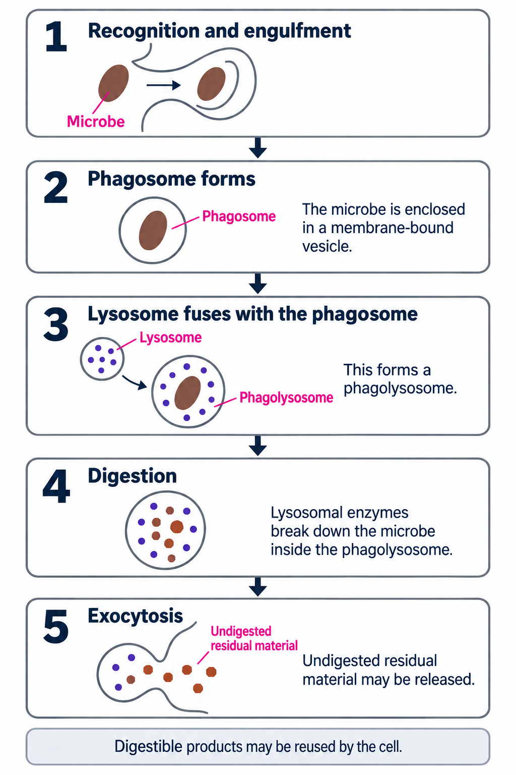
c. Phagocytic cells (macrophage)
Phagocytic white blood cells | Functions and features |
Neutrophils | • Engulf microorganism or foreign particles |
Monocytes | • Become macrophages for phagocytic activity |
Eosinophils | • Minor phagocytic activity • Release enzymes to kill parasites • Discharge toxins from enzyme damage to tissues |
d. Natural killer cells
- Release molecules (perforin) to kill infected or tumor cells.
- Apoptosis – programmed cell death.
D. Specific immunity
– the primary adaptive response develops more slowly at first; memory responses to the same antigen are typically faster and stronger.
1. Basic theory
a. Terminology
- Antibiotic
– A drug that kills bacteria or inhibits their growth.
– Antibiotics act through several mechanisms, including inhibition of bacterial cell-wall synthesis or protein synthesis.
– Antibiotics do not treat viral infections.
- Cytokines
– proteins secreted from immune cells.
– function to help activate lymphocytes and coordinate the immune system against infection.
b. Structures of B cell and antigen
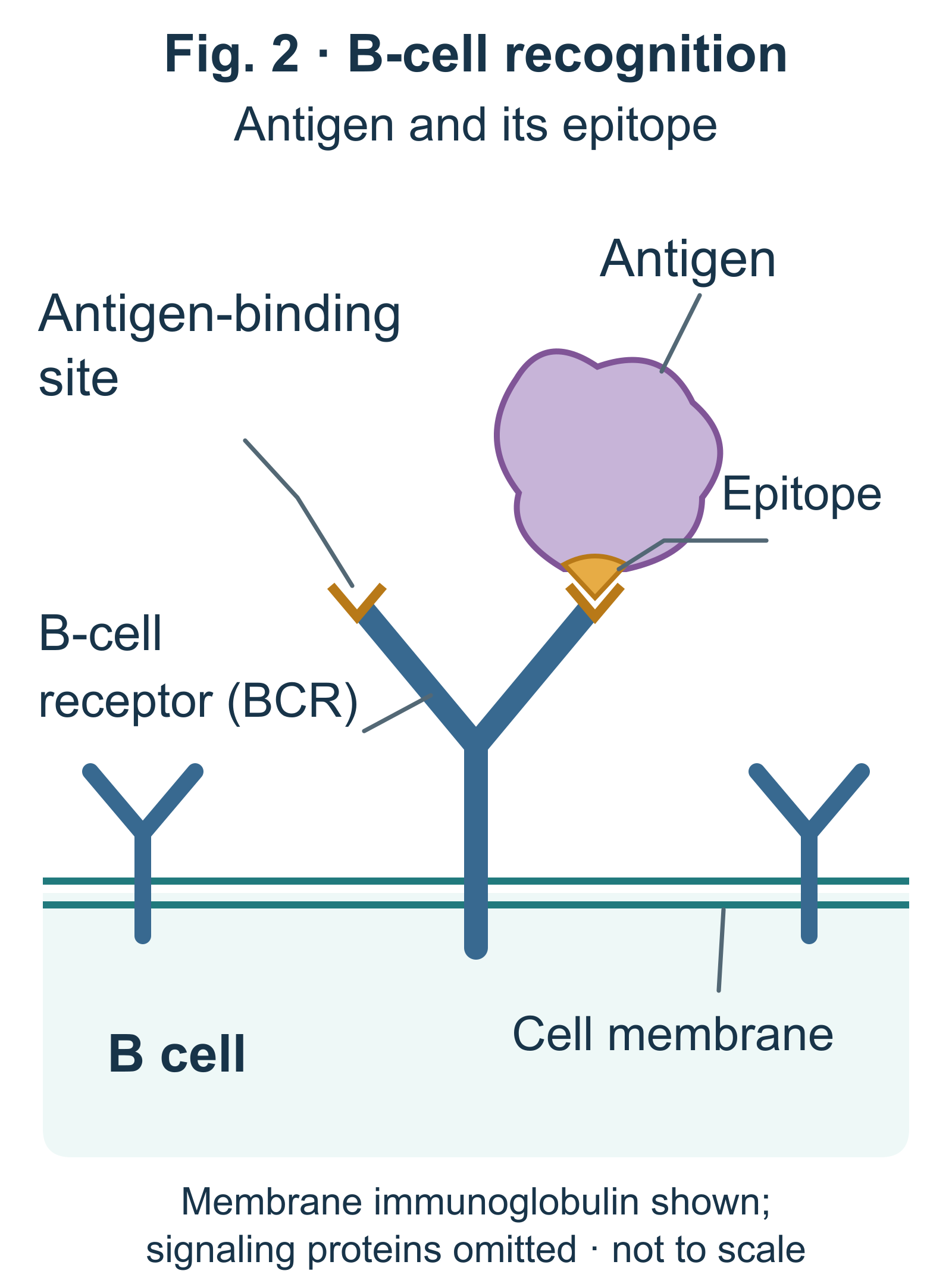
- Antibodies
– Antibodies are immunoglobulins secreted primarily by differentiated plasma cells and found in blood plasma and interstitial fluid.
– Membrane-bound immunoglobulin on B cells functions as the B-cell receptor (BCR).
– function as identifying and binding foreign microbes for destruction.
- Antigen
– foreign molecules or microbes detected by lymphocytes.
- Epitope
– antigenic determinants.
– recognition of site of antigens.
c. Lymphocytes
- The major lymphocyte lineages include B cells, T cells, and natural killer (NK) cells.
- Major function as body’s defenses
d. Structure of B cell and T cell receptors
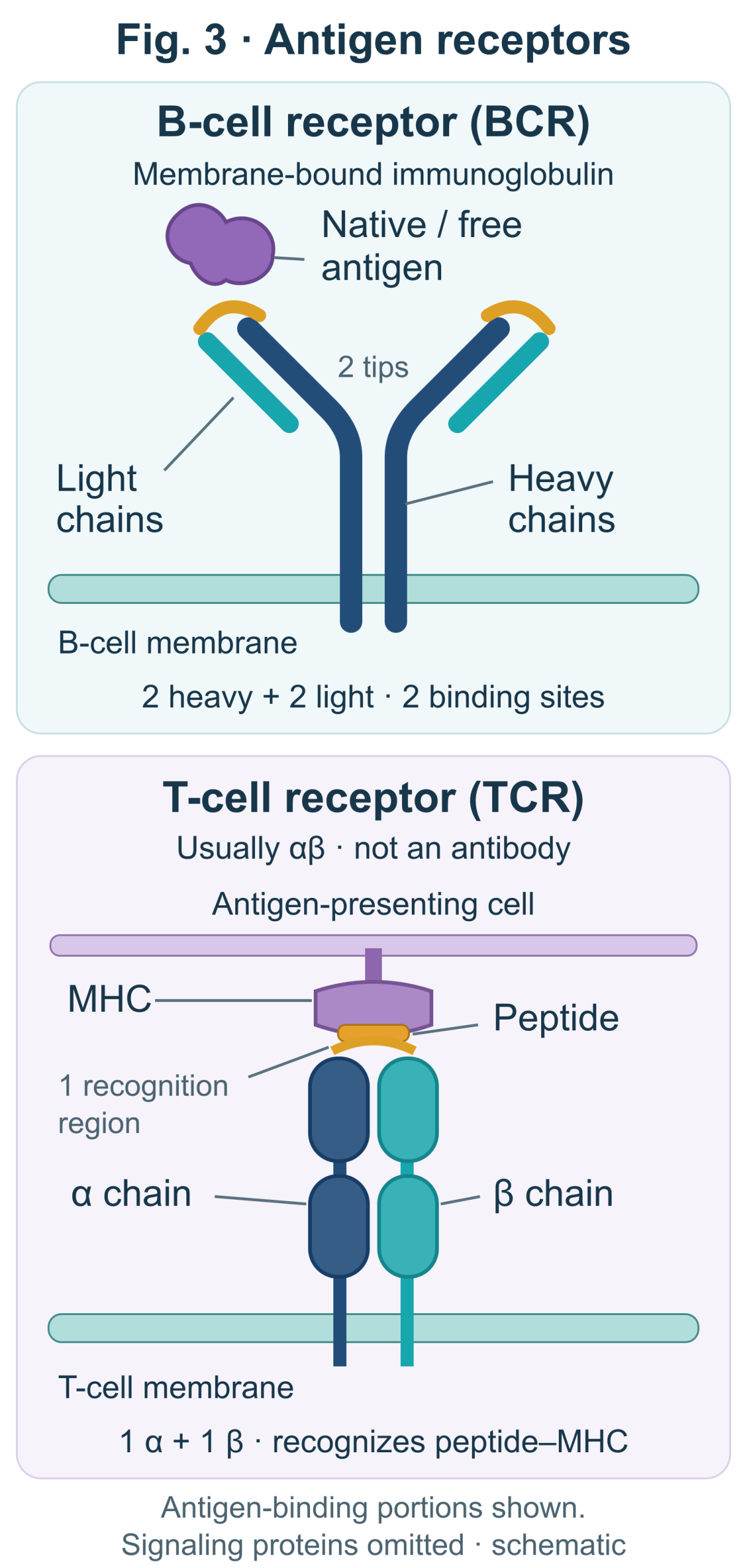
Receptors | Features |
B cell receptor | • Y shape with four polypeptide chains (two heavy chains and two light chains) • A B-cell receptor contains two heavy and two light immunoglobulin chains linked by disulfide bonds. |
T cell receptor | • Two lines shape with two different polypeptide chains (α and β chains) • Disulfide bridge links α and β chains |
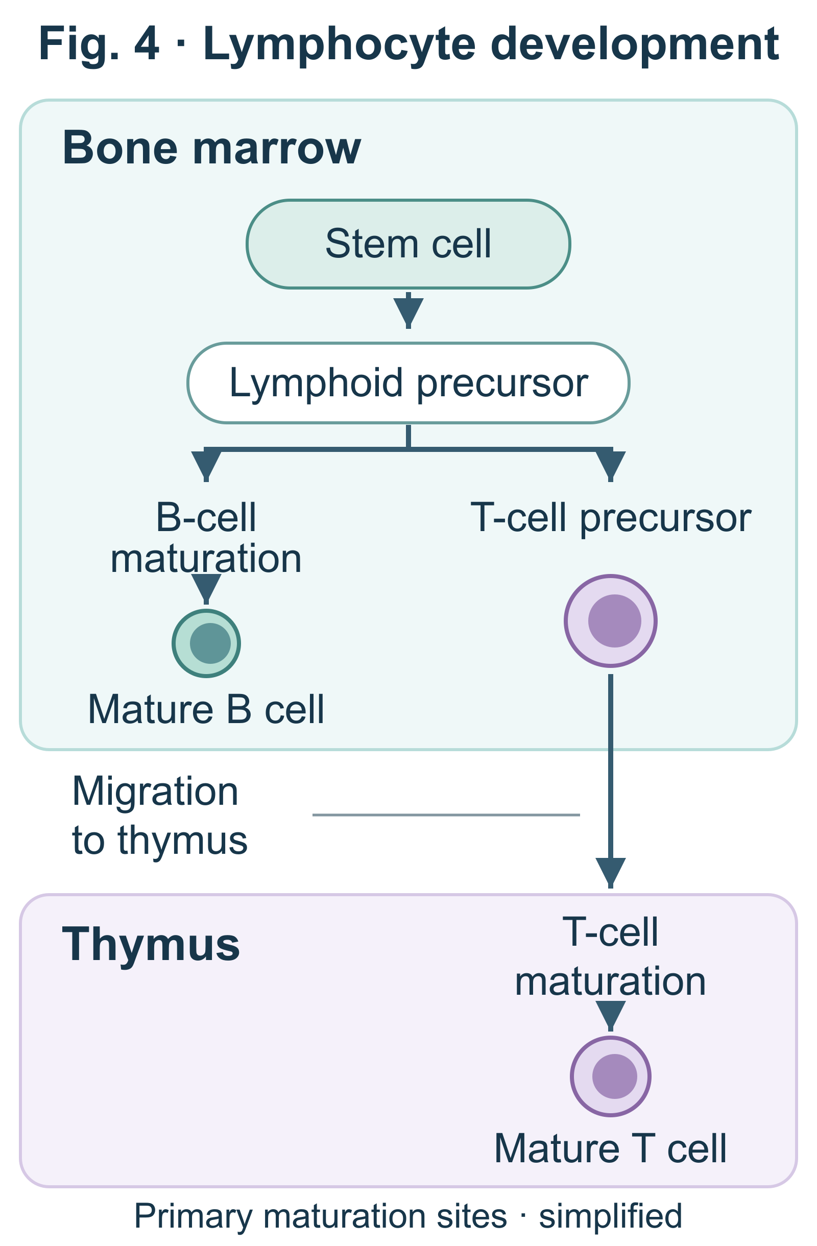
e. Lymphocyte development
: Lymphocytes arise from stem cells in the bone marrow. B cells mature in the bone marrow, whereas T-cell precursors migrate to the thymus and mature there.
f. Clonal selection of lymphocyte
- Clonal selection – antigen driven activation of lymphocytes
- B-cell effector cells (plasma cells)
– Activated B cells differentiate into antibody-secreting plasma cells; some are short-lived, whereas others are long-lived.
– Secrete antibodies specific for the antigen that activated the B-cell clone.
- Memory cells
– long-lived cells.
– bearing receptors specific for the same antigen.
- Primary immune response
– Activated B cells proliferate and differentiate into antibody-secreting plasma cells; CD8 T cells can become cytotoxic effector cells.
– Illness may occur while the primary immune response develops, but it is not inevitable.
- Secondary immune response
– much faster immune response from same antigen, more prolonged, and greater magnitude than primary immune response.
2. Humoral and cell-mediated immunities
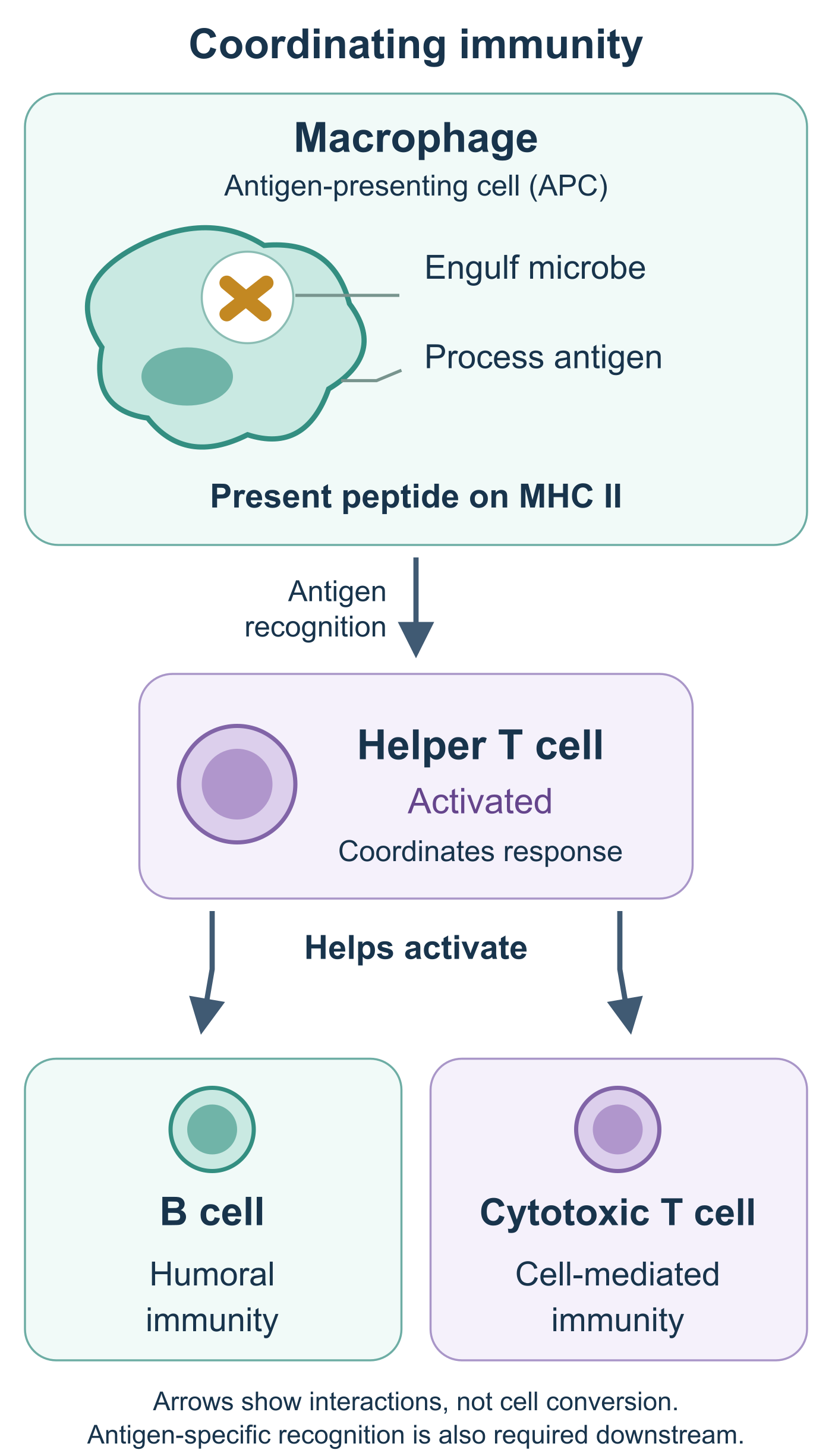
- Macrophage
– engulfs microorganisms and presents antigens.
- Helper T cell
– responds to antigens, which are displayed from macrophages.
– stimulates the activation of B cells and cytotoxic T cells.
a. Humoral immunity
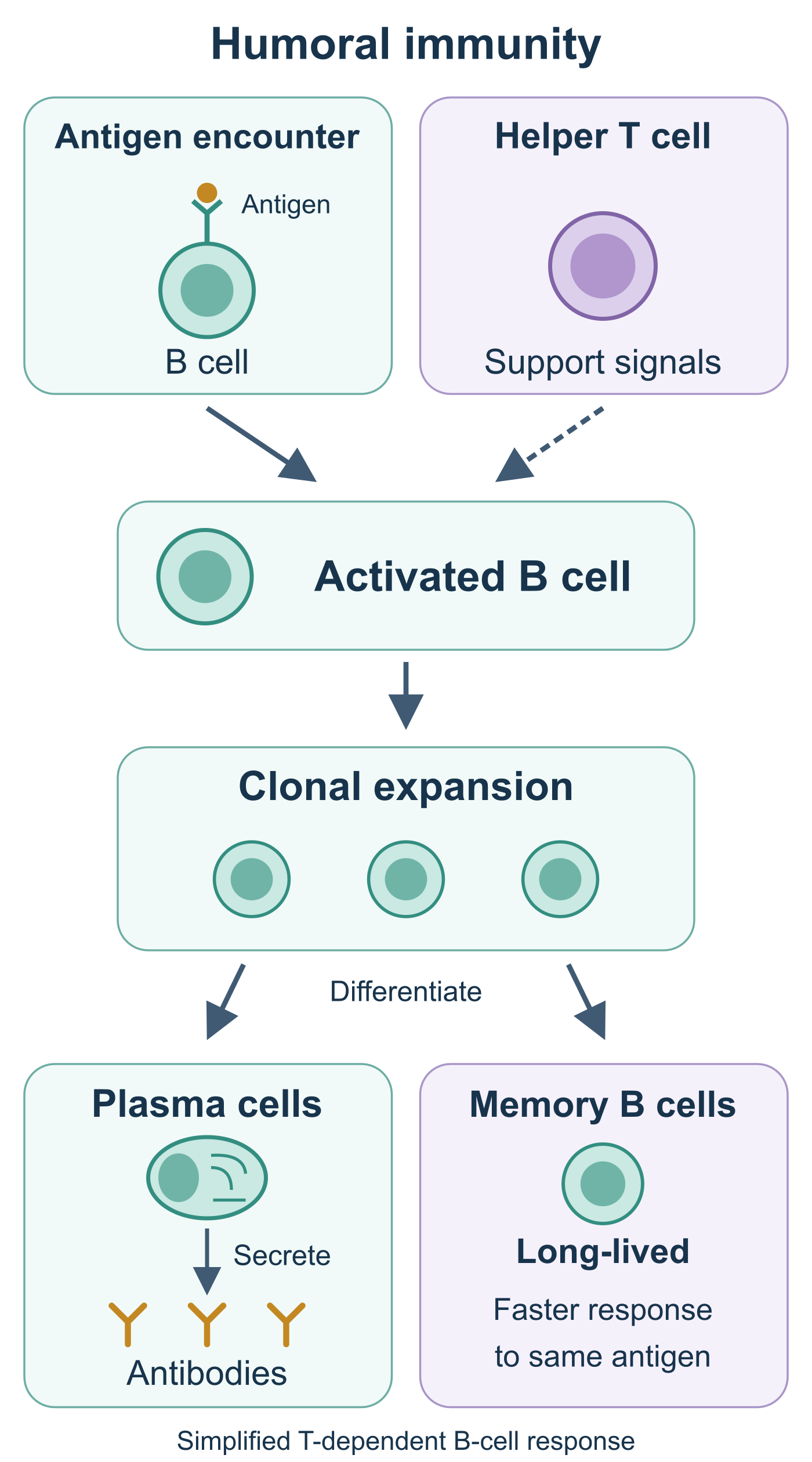
: activates B cells to proliferate and differentiate into plasma cells, which secrete antibodies that help defend against pathogens in the blood and lymph.
- B Cells
– important role in the humoral immune response.
– respond to pathogens and are activated by helper T cell.
– make the clone of memory B cells.
– Activated B cells proliferate and differentiate into plasma cells that secrete antibodies.
- Memory B cells
- Plasma cells – produce antibodies
- Classes of antibodies
Classes | Shapes | Features |
IgA |
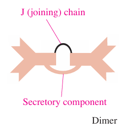
| • Important role in newborn immunity • Present in breast milk, mucosal surfaces, tears, and saliva • Serve localized defense of mucous membranes |
IgD |
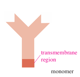
| • Important antigen receptor on B cells • Antigen receptor and differentiation of B cells |
IgE |
| • Involved in allergic reaction |
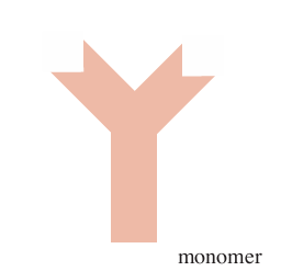
IgG |
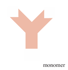
| • Most common antibody in blood • Serve for neutralization, agglutination of antigens, and opsonization • Important role in fetus and newborn immunity • Cross the placenta • Often prominent in secondary immune responses and in later phases of class-switched responses. |
IgM |
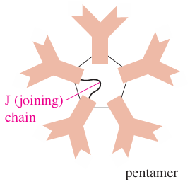
| • Often prominent early in a primary immune response. • Useful in complement activation • Serve for neutralization and agglutination of antigens |
b. Cell-mediated immunity
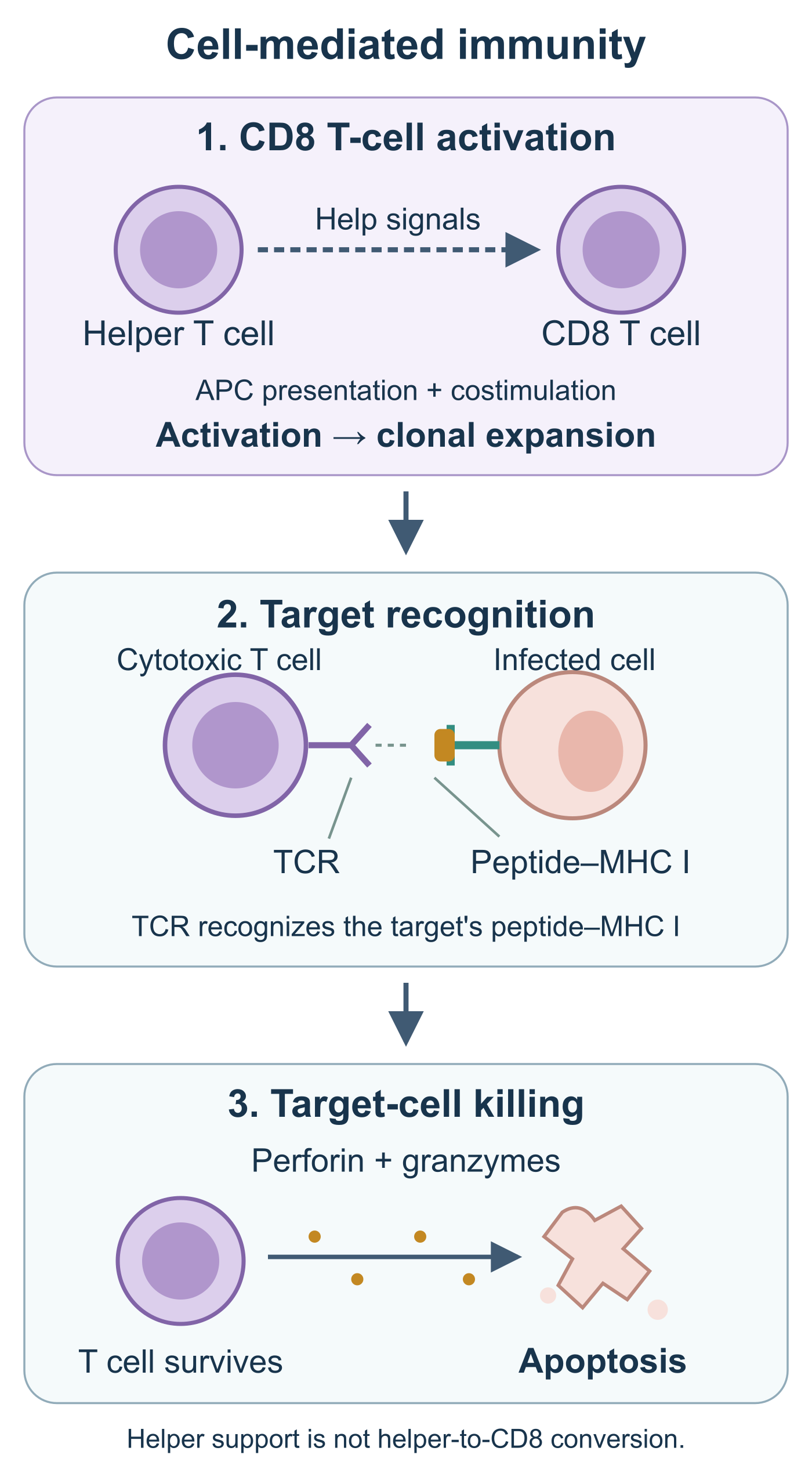
: activates the clone of cytotoxic T cells, which destroy cancer cells or infected cells.
- Cytotoxic T cell
– important role in the cell-mediated immune response.
– T cells mature in the thymus into distinct CD4 and CD8 lineages. After activation, CD8 T cells can differentiate into cytotoxic effector T cells.
– uses the protein perforin to destroy cancer cells or infected cells.
- Class II MHC molecules
– MHC class II molecules are expressed primarily by professional antigen-presenting cells, including dendritic cells, macrophages and B cells, and present peptide antigens to CD4 helper T cells.
- Class I MHC molecules
– MHC class I molecules are expressed on nearly all nucleated cells and present intracellular peptides to CD8 T cells.
- T cell receptor
– A typical alpha-beta T-cell receptor recognizes peptide antigen presented by an MHC molecule, rather than free antigen.
E. Immune responses
1. Generating immunity
- Active immunity
– Active immunity develops when the individual’s own immune system responds to antigen exposure through infection or vaccination.
– eg/ Vaccination
- Passive immunity
– Passive immunity is protection provided by antibodies received from another source.
– Maternal IgG crosses the placenta to the fetus. IgA is transferred mainly through breast milk and helps protect the infant’s mucosal surfaces.
Rh-related hemolytic disease: in a sensitized Rh-negative mother, anti-D IgG can cross the placenta and damage an Rh-positive fetus’s red blood cells. This is antibody-mediated disease, not protective passive immunity.
2. Blood group
ABO red blood cell transfusion compatibility only; Rh factor, other blood-group antigens, and crossmatching must also be considered.
Blood type | Antibodies | Reaction to other blood type | Possible transfusion blood type from others |
A | Anti-B | B, AB | A, O |
B | Anti-A | A, AB | B, O |
AB | None | None | A, B, AB, O |
O | Anti-A, Anti-B | A, B, AB | O |